Definition
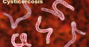
Opisthorchiasis is an infection caused by either of the two parasite worms, the cat liver fluke, or scientifically called Opisthorchis felineus (also known as Opisthorchis tenuicollis), or the Southeast Asian liver fluke (called as Opisthorchis viverrini). The term “fluke” means flatfish, since these parasites are flat.
Opistorhoz in fish
History
Major texts state that the human symptoms of the disease Opisthorchiasis was first described in 1875. In 1884, the parasite Opisthorchis felineus was first discovered in cat liver in Northern Italy. In 1891 a Russian scientist, K. N. Vinogradov discovered this parasite in a human and named it the “Siberian liver fluke.”
Opisthorchis viverrini is the only parasite that causes Opisthorchiasis in the heavily infected area of Thailand. The first case of Opisthorchiasis in Thailand was discovered in 1911 through autopsy.
Epidemiology
Opisthorchiasis is prevalent where raw cyprinid fishes are a staple of the diet. Prevalence rises with age; children under the age of 5 years are rarely infected by Opisthorchis viverrini. Males may be affected more than females. The WHO estimates that foodborne trematodiases (infection by worms or “flukes”, mainly Clonorchis, Opisthorchis, Fasciola and Paragonimus species) affect 56 million people worldwide and 750 million are at risk of infection. Eighty million are at risk of opisthorchiasis, 67 million from infection with Opisthorchis viverrini in Southeast Asia and 13 million from Opisthorchis felineus in Kazakhstan, Russia including Siberia, and Ukraine. In the lower Mekong River basin, the disease is highly endemic, and more so in lowlands, with a prevalence up to 60% in some areas of northeast Thailand. However, estimates using newer polymerase chain reaction-based diagnostic techniques indicate that prevalence is probably grossly underestimated. In one study from the 1980s, a prevalence of over 90% was found in persons greater than 10 years old in a small village near Khon Kaen in northeast Thailand in the region known as Isaan. Sporadic cases have been reported in case reports from Malaysia, Singapore, and the Philippines. Although overall prevalence declined after initial surveys in the 1950s, increases since the 1990s in some areas seem associated with large increases in aquaculture.
Risk factors of Opisthorchiasis
The risk factors of Opisthorchiasis include:
- People who eat freshwater fish that is raw, undercooked, pickled, or unprocessed (even if imported), containing the parasitic worms are at a high risk of contracting Opisthorchiasis.
- Living near freshwater water bodies, such as rivers, ponds, or lakes, in the endemic regions of Eastern Europe, Russia, Thailand or Laos where these parasites are highly prevalent and consuming raw fish
It is important to note that having a risk factor does not mean that one will get the condition. A risk factor increases ones chances of getting a condition compared to an individual without the risk factors. Some risk factors are more important than others.
Causes
The causes of Opisthorchiasis include:
- Opisthorchiasis is a foodborne infection caused by the cat liver fluke or the Southeast Asian liver fluke, a parasitic worm which grows and resides in the bile ducts of the liver in humans and fish-eating mammals. It is acquired by eating freshwater fish containing the fluke larvae
- The eggs of Opisthorchis are taken in by snails that live in the freshwaters. These eggs hatch and the infected snails release microscopic larvae that enter freshwater fish (which serves as an intermediate host). Humans (or mammals), who are the final hosts, are infected when they eat raw or undercooked fish containing the cysts
- On entering the human body, the cyst travels to the intestine and then moves to the liver and resides in the bile ducts, where they grow into adult worms. The adult worm lays and hatches eggs that is passed on to the intestine and then into the feces
- Opisthorchis felineus is usually a parasite of cats, dogs, pigs, and whales, while Opisthorchis viverrini is commonly found in dogs and cats
Life cycle of Opisthorchiasis
Life cycle of fluke
The adult flukes deposit fully developed eggs that are passed in the feces.
- After ingestion by a suitable snail (first intermediate host)
- The eggs release miracidiaThe number
2(a) Which undergo in the snail several developmental stages (sporocysts
2(b) RediaeThe number
2(c) CercariaeThe number
2(d) Cercariae are released from the snail
- And penetrate freshwater fish (second intermediate host), encysting as metacercariae in the muscles or under the scales
- The mammalian definitive host (cats, dogs, and various fish-eating mammals including humans) become infected by ingesting undercooked fish containing metacercariae. After ingestion, the metacercariae excyst in the duodenum
- And ascend through the ampulla of Vater into the biliary ducts, where they attach and develop into adults, which lay eggs after 3 to 4 weeks
- The adult flukes (O. viverrini: 5 mm to 10 mm by 1 mm to 2 mm; O. felineus: 7 mm to 12 mm by 2 mm to 3 mm) reside in the biliary and pancreatic ducts of the mammalian host, where they attach to the mucosa.
Microscopic view of Opisthorchis viverrini
Symptoms
Most individuals infected with Opisthorchiasis do not present any symptoms. Individuals infected for a long time (chronic) may get serious illness. A long-standing inflammation or re-infection of the bile ducts could lead to scarring of the bile duct, and inflammation of the liver.
The signs and symptoms of Opisthorchiasis may include:
- Abdominal pain, nausea, and diarrhea
- Any pathology (infection) of the bile duct and adjacent organs can cause abdominal pain (particularly under the rib cage to the right), jaundice, fever, or chills
- Biliary stone formation (cholelithiasis): Inflammation and scarring of the bile ducts leads to stasis (decreased flow) of bile, which may to lead to the formation of stones
- Due to bile stones and narrowing changes, the affected individuals may get the following:
- Jaundice
- Intermittent biliary duct obstruction can cause abdominal pain (biliary colic)
- Cholangitis – infection and inflammation of the bile duct
- Cholecystitis – infection and inflammation of the gall bladder
- Pancreatitis (inflammation of the pancreas) due to obstruction of the tube (duct) that is common to the liver bile duct and pancreas
Complications of Opisthorchiasis
The complications of Opisthorchiasis could include:
- Scarring of the bile duct or gallstone formation that could lead to diseases such as
- Cholecystitis (infection of the gallbladder)
- Cholangitis (infection of the biliary duct)
- Hepatitis (inflammation of the liver)
- Cholangiocarcinoma – cancerous growth in one of the duct that carries bile from the liver to the small intestines
- Researchers believe that O. viverrini (Southeast Asian liver fluke) can cause cholangiocarcinoma, especially in the presence of dietary nitrosamines (which is used as a preservative).
Diagnosis and test
The following tests and procedures may be used to diagnose Opisthorchiasis:
- Thorough evaluation of the individual’s medical history and a complete physical examination, especially of the abdomen
- During history taking the physician may want to know the following:
- When the symptoms began and whether they are becoming worse
- Information on dietary habits (especially raw fish consumption), travel history, and places of residence
- The diagnosis of Opisthorchiasis or clonorchiasis (a similar condition) is suspected when the individual is accustomed to consuming raw fish and has signs and symptoms pertaining to the right side of the abdomen
- Consultation with a gastroenterologist and or infectious disease specialist may be necessary
- Blood tests:
- Complete blood count may show eosinophilia – increased levels of white blood cell eosinophils in the peripheral blood. It is usually seen with parasitic infections
- Tests for checking antibody levels for the parasite
- Examination of a stool sample may be undertaken to detect the presence of the eggs of Opisthorchis to confirm diagnosis
- Imaging tests may be performed to look for disease activity in the bile duct, liver, gallbladder, or pancreatic duct
- Ultrasound, CT, or MRI scans of the liver, gallbladder, biliary and pancreatic ducts may be taken
- Endoscopic retrograde cholangiopancreatography (ERCP) may be done to check for abnormalities in the biliary and pancreatic ducts
- Many clinical conditions may have similar signs and symptoms. Your healthcare provider may perform additional tests to rule out other clinical conditions to arrive at a definitive diagnosis.
Treatment and medications
The treatment options of Opisthorchiasis include:
- The World Health Organization (WHO) recommends anti-worm medication praziquantel for infected individuals. Praziquantel may be given to individuals as a preventative measure; to individuals who have no signs of the disease, but live in the endemic regions
- Surgery may be required, if obstruction of the bile duct persists, despite treatment with praziquantel to remove the worms and/or stones
- Treatment of complications as required with intravenous antibiotics and through surgical intervention
Prevention of Opisthorchiasis
- Proper cooking of fish..
- Freezing fish intended for raw consumption.
- Use of molluscicides is the most frequent public health intervention, as it prevents the transmission of many other trematodes, including Schistosoma spp.
- Treatment of animals to reduce the reservoir and stock losses has been used.
- Prophylactic use of praziquantel.


can I go to private hospital for the treatment of cholethiasis
Kindly consult a gastroenterologist for treatment.