Definition
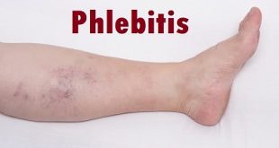
Peripheral vascular disease (PVD) is a problem with poor blood flow. It affects blood vessels outside of the heart and brain and gets worse over time. Parts of the body, like the brain, heart, arms, or legs, may not get enough blood. The legs and feet are most commonly affected. Other blood vessel problems like deep vein thrombosis (DVT), varicose veins, and chronic venous insufficiency are linked to PVD.
PVD is often found in people with problems with the arteries that supply blood to the heart (coronary artery disease). That is because atherosclerosis, which causes coronary artery disease, affects arteries throughout the body. Atherosclerosis is the buildup of plaque on the walls of the arteries. Plaque is made up of cholesterol, fat, and waste from the cells, and other materials. It causes the arteries to become stiff and narrow.
Epidemiology
Peripheral arterial disease (PAD) affects approximately 30% of older individuals in the general population. The principal symptom, intermittent claudication, occurs only rarely in those under 50 years of age, but increases dramatically in older individuals.
Types of Peripheral Vascular Disease
The two main types of PVD are functional and organic PVD.
- Functional PVD means there’s no physical damage to your blood vessels’ structure. Instead, your vessels widen and narrow in response other factors like brain signals and temperature changes. The narrowing causes blood flow to decrease.
- Organic PVD involves changes in blood vessel structure like inflammation, plaques, and tissue damage.
Risk factors
Risk factors contributing to PVD are the same as those for atherosclerosis:
- Smoking- Tobacco use in any form is the single most important modifiable cause of PVD internationally. Smokers have up to a tenfold increase in relative risk for PVD in a dose-related effect. Exposure to second-hand smoke from environmental exposure has also been shown to promote changes in blood vessel lining (endothelium) which is a precursor to atherosclerosis.
- Diabetes mellitus – Causes between two and four times increased risk of PVD by causing endothelial and smooth muscle cell dysfunction in peripheral arteries. Diabetics account for up to 70% of nontraumatic amputations performed, and a known diabetic who smokes runs an approximately 30% risk of amputation within 5 years.
- Hypertension – Elevated blood pressure is correlated with an increase in the risk of developing PVD, as well as in associated coronary and cerebrovascular events (heart attack and stroke).
- Risk of PVD also increases in individuals who are over the age of 50, male, obese, or with a family history of vascular disease, heart attack, or stroke.
- Other risk factors which are being studied include levels of various inflammatory mediators such as C-reactive protein, homocysteine.
Causes of Peripheral Vascular Disease
The most common cause of PVD is peripheral artery disease, which is due to atherosclerosis. Fatty material builds up inside the arteries and mixes with calcium, scar tissue, and other substances. The mixture hardens slightly, forming plaques. These plaques block, narrow, or weaken the artery walls. Blood flowing through the arteries can be restricted or completely blocked.
Other causes of PVD include:
Blood clots: A blood clot can block a blood vessel.
Diabetes: The high blood sugar level present with diabetes can, over time, damage blood vessels. This makes them more likely to become narrow or to weaken. People with diabetes often also have high blood pressure and a high level of fats in the blood. Both conditions can accelerate the development of atherosclerosis.
Inflammation of the arteries or arteritis: Arteritis can cause narrowing or weakening of the arteries. Some autoimmune conditions lead to vasculitis. The inflammation can affect not just arteries, but other organ systems too.
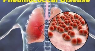
Infection: The inflammation and scarring caused by infection can block, narrow, or weaken blood vessels. Both salmonellosis (infection with Salmonella bacteria) and syphilis are two infections traditionally known to infect and damage blood vessels.
Structural defects: Defects in the structure of a blood vessel can cause narrowing. Most of these are acquired at birth, and the cause is unknown. Takayasu disease is a vascular disease that damages the aorta, the large blood vessel carrying blood from the heart to the body. It is most common among females of Asian origin.
Injury: Blood vessels can be injured in an accident such as a car wreck or a bad fall.
Peripheral Vascular Disease Symptoms
About half the people diagnosed with PVD are symptom free. For those with symptoms, the most common first symptom is painful leg cramping that occurs with exercise and is relieved by rest (intermittent claudication). During rest, the muscles need less blood flow, so the pain disappears. It may occur in one or both legs depending on the location of the clogged or narrowed artery.
Other symptoms of PVD may include:
- Changes in the skin, including decreased skin temperature, or thin, brittle, shiny skin on the legs and feet
- Weak pulses in the legs and the feet
- Gangrene (dead tissue due to lack of blood flow)
- Hair loss on the legs
- Impotence
- Wounds that won’t heal over pressure points, such as heels or ankles
- Numbness, weakness, or heaviness in muscles
- Pain (described as burning or aching) at rest, commonly in the toes and at night while lying flat
- Paleness when the legs are elevated
- Reddish-blue discoloration of the extremities
- Restricted mobility
- Severe pain when the artery is very narrow or blocked
- Thickened, opaque toenails
The symptoms of PVD may look like other conditions. See your healthcare provider for a diagnosis.
Peripheral Vascular Disease Complications
Complications of peripheral vascular disease most often occur because of decreased or absent blood flow. Such complications may include:
- Amputation (loss of a limb)
- Heart attack
- Poor wound healing
- Restricted mobility due to pain or discomfort with exertion
- Severe pain in the affected extremity
- Stroke (three times more likely in persons with PVD)
By following an aggressive treatment plan for peripheral vascular disease, complications such as these may be prevented.
Diagnosis and test
To diagnose PVD, your health care provider will do a complete medical history and physical exam. Other tests may include:
Angiography: A procedure that allows the health care provider to see blocked or narrowed blood vessels. This procedure involves inserting a thin, flexible tube into an artery in the leg and injecting a contrast dye. The contrast dye makes the arteries and veins visible on the X-ray.
Ankle-brachial index (ABI): An ABI is a comparison of the blood pressure in the ankle with the blood pressure in the arm. It helps to diagnose PVD and checks for changes in blood flow over time.
Doppler ultrasound flow studies: This uses high-frequency sound waves and a computer to create images of blood vessels, tissues, and organs. Doppler technique is used to measure and assess the flow of blood. Faintness or absence of sound may mean an obstruction in the blood flow.
Magnetic resonance angiography (MRA): This procedure uses large magnets, radio frequencies, and a computer to produce detailed images. An MRA is typically only done if plans are being made to restore normal blood flow through a procedure or surgery.
Photoplethysmography (PPG): This exam is like the ankle brachial index except that it uses a very tiny blood pressure cuff around the toe and a PPG sensor (infrared light to evaluate blood flow near the surface of the skin) to record waveforms and blood pressure measurements. These measurements are then compared to the systolic blood pressure in the arm.
Pulse volume recording (PVR) waveform analysis: This test checks for changes in the amount of blood in the legs.
Segmental blood pressure measurements: Blood pressure measurements are taken at various points along the arms or legs. The level and severity of disease can be identified through segmental blood pressures.
Exercise tests: Some of the above tests can be done before and after exercise. This is especially helpful for evaluating patients with symptoms during exercise.
Treatment and medications
Treatment options may include:
Medications- To help treat atherosclerosis, such as statins to lower LDL cholesterol and antihypertensive drugs to lower blood pressure.
Drugs to treat blood clots- Ttreatment may include various medications (including anticoagulants and anti-platelet drugs) to prevent blood clots from developing and medications (including thrombolytics) that dissolve existing blood clots.
Angioplasty- This procedure, usually performed under sedation and local anaesthetic, involves threading a thin tube (catheter) into the narrowed blood vessel through a small incision, usually in the leg. Once the catheter reaches the narrowed or blocked site, the small balloon on its tip is inflated. This widens the blood vessel and improves blood flow. Angioplasty is usually considered as a temporary measure.
Surgical insertion of a stent- A stent is a metal ‘sleeve’ that is implanted inside the narrowed blood vessel during an angioplasty procedure to prop it open. Stents may be impregnated with medications that help to prevent scar tissue from narrowing the treated area of blood vessel.
Atherectomy- This operation involves cutting away the fatty obstruction with a small scalpel-like instrument.
Bypass surgery- This operation is usually only considered in severe cases that don’t respond to other treatments or in cases that involve large sections of the diseased blood vessel. A section of healthy vein is taken from somewhere else in the body and surgically grafted to re-route blood flow around the blockage in the affected blood vessel. A surgeon may sometimes use a piece of synthetic tubing to detour blood flow.
Prevention of Peripheral Vascular Disease
You can reduce your risk of developing PVD through a healthy lifestyle. This includes:
- Avoiding smoking
- Controlling your blood sugar, if you have diabetes
- Setting an exercise goal of 30 minutes a day, five times a week
- Working to lower cholesterol and blood pressure
- Eating a healthy diet that’s low in saturated fat
- Keeping your weight at a healthy level
Talk to your doctor if you experience symptoms of PVD. Early diagnosis can help you and your doctor find ways to reduce your symptoms and increase the effectiveness of your treatment.


Can you recommend some on the counter medication? I’ve very painful leg cuffs from the knee to the anckle and under the toes. So weak I can’t stand
It would be better to consult the doctor to get the prescribed medicines.