Description
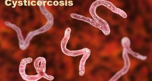
Neurocysticercosis, the infection caused by the larval form of the tapeworm Taenia solium, is the most common parasitic disease of the central nervous system and the most common cause of acquired epilepsy worldwide. The larval cysts can infect various parts of the body causing a condition known as cysticercosis. Larval cysts in the brain cause a form of cysticercosis called neurocysticercosis which can lead to seizures.
Taenia solium is a two-host zoonotic cestode. The adult stage is a 2-4m-long tapeworm that lives in the small intestine of humans. No other final hosts are known for T. solium tapeworms in nature. As in all cestodes, the gravid proglottids at the terminal end of the worm are full of eggs that are the source of infection with the larval stage, or cysticercosis. The natural intermediate host is the pig, harboring larval cysts anywhere in its body.
Causes and Transmission of Neurocysticercosis
Cysticercosis in humans is acquired by ingesting eggs of the pork tapeworm (T. solium). It is not from ingestion of undercooked meat. Eggs are found only in human feces, and a carrier can shed eggs into the environment in a stool or transmit them on unwashed hands and fingernails, to other people or to pigs, who are infected when they swallow contaminated soil, water or food. These eggs can live for 2 months in water, soil or vegetation.
Once ingested, the eggs hatch in the small intestine and the larvae migrate to various tissues throughout the body, where they form cysts. This is called cysticercosis. If the cysts are in the brain, the condition is called neurocysticercosis, the most severe form of the disease.
Stages of Neurocysticercosis
There are four main stages (also known as Escobar’s pathological stages):
- Vesicular: viable parasite with intact membrane and therefore no host reaction.
- Colloidal vesicular: parasite dies within 4-5 years untreated, or earlier with treatment and the cyst fluid becomes turbid. As the membrane becomes leaky edema surrounds the cyst. This is the most symptomatic stage.
- Granular nodular: edema decreases as the cyst retract further; enhancement persists.
- Nodular calcified: end-stage quiescent calcified cyst remnant; no edema.
Risk factors
People are at a higher risk for getting neurocysticercosis by swallowing parasite eggs if they:
- Have a pork tapeworm infection (this is called autoinfection)
- Live in a household with someone who has a pork tapeworm
- Eat food made by someone with a pork tapeworm infection
Symptoms
Clinical manifestations of neurocysticercosis vary with the locations of the lesions, the number of parasites, and the host’s immune response. There is a variable time interval between the point of infection and the onset of symptoms (ranging from 1-30 years). Many patients are asymptomatic. Possible symptomatic presentations include the following:
- Epilepsy
- A headache
- Dizziness
- Stroke
- Neuropsychiatric dysfunction
- The onset of most symptoms is usually subacute to chronic, but seizures present acutely
Abnormal physical findings, which occur in 20% or less of patients with neurocysticercosis, depend on where the cyst is located in the nervous system and include the following:
- Cognitive decline
- Dysarthria
- Extraocular movement palsy or paresis
- Hemiparesis or hemiplegia, which may be related to stroke, or Todd paralysis
- Hemisensory loss
- Movement disorders
- Hyper/hyporeflexia
- Gait disturbances
- Meningeal signs
Complications of Neurocysticercosis
In untreated or late diagnosed cases of Neurocysticercosis, the following complications are seen:
- Hydrocephalous
- Large cysts
- Multiple lesions in the brain along with edema
- Chronic meningitis
- Vasculitis
- Complications also include paralysis, partial blindness, lost ability to speak and some go into coma.
Most of these complications of Neurocysticercosis do not respond very well to the treatment and may lead to poor prognosis.
Diagnosis of Neurocysticercosis
The doctor will perform the two tests of the brain
- Magnetic Resonance Imaging (MRI) Scan to Diagnose Neurocysticercosis: MRI scan of the brain shows whitish bobs on brain image which indicate the cysts of tapeworms.
- Computed Tomography (CT) Scan: CT scan is used for detection of calcifications.
MRI image showing cysts in the brain
When just MRI and CT scan results are not sufficient additional tests are required as:
- ELISA of Cerebrospinal fluid (CSF) is done
- Cerebrospinal fluid or CSF analysis for the number of mononuclear cells and eosinophils, glucose levels, protein levels, and IgG antibody levels are done
- Stool examination
- Brain biopsy is done in extreme cases
Treatment of Neurocysticercosis
The treatment of neurocysticercosis depends on the life stage of the cyst and its complications. It goes as follows:
If the parasite is dead, then the treatment for Neurocysticercosis is directed against the symptoms.
- Anticonvulsants are used to treat the seizures and the duration of this treatment varies.
- If parenchymal lesions resolve without calcifying and patient remains seizure-free, then anticonvulsant therapy may be discontinued after 2 years.
- The First Line /classical anticonvulsant medications include: Phenytoin Carbamazepine, Phenobarbitals
- Second line / Newer medicines: Valproic acid, Lamotrigine, Levetiracetam, Zonisamide.
If the parasite is alive and active then,
- Immunosuppressants are administered followed by anticysticercal drugs or anticystocisal drugs such as praziquantel and albendazole.
- Antiparasitic drug treatment with albendazole is useful in the racemose type of cysticercosis.
- Similarly, for giant subarachnoid cysts, multiple anticysticeral drug treatment is required.
- If the patient has encephalitis then steroids are given before use of anticysticeral drugs.
Surgical treatment for Neurocysticercosis is given when –
- Hydrocephalus is present due to ventricular cysts. The excess fluid accumulation exerts pressure and requires shunt for its removal.
- Multiple cysts in racemose form present in the subarachnoidal space
- Obstruction is noticed due to arachnoiditis.
Since Neurocysticercosis is a complex disease, hence there is no single treatment pattern for all patients. Overall, the patient status, number, and location of parasites determine the type of treatment to be given.
Prevention
Neurocysticercosis is the leading cause of epilepsy in the developing world. Infection from this tapeworm is preventable. Following measures can be used to prevent it.
- To avoid it, people should be sure the meat they eat is cooked completely hygiene is important
- Involves washing hands thoroughly after using the bathroom
- Proper sewage disposal and cattle should be kept away from the house.
- Health education campaigns are effective in the prevention and control of many infectious diseases. Participation of the community and schools in maintaining hygienic and sanitary conditions.
- Screening of farm workers for taeniosis, and treatment if warranted.
- Control of pig and cattle marketing systems, including the provision of incentives to ensure owner compliance
- Health education to both farmer and consumer, especially on the cooking of meat.
- Meat inspection to prevent infection.
- Improved farm management to ensure that pigs and cattle are protected from ingesting feed or water contaminated with human feces to prevent Cysticercosis in animals.


very nice
nice information…. thanks a lot
very helpful…
is there any treatment for those parasites that caused the problem in the brain for the seizures
the seizure can be treated with normal drugs (Acetazolamide, Carbamazepine) but to remove parasites surgery is needed.
thanks for the information,my daughter 22years old was on anticonvulsant drugs for one year but the convulsions continued but after she stopped it can occur after 3month,but the big problem now is headache every time side ways, what could be the problem.
Please consult a doctor to get rid of this problem.