Nephrocalcinosis – Summary
Nephrocalcinosis is a disorder that occurs when too much calcium is deposited in the kidneys. It commonly occurs in premature infants. Individuals may not have symptoms or may have symptoms related to the condition causing Nephrocalcinosis. If kidney stones are present, symptoms may include blood in the urine, fever and chills, nausea and vomiting, and severe pain in the belly area, sides of the back (flank), groin, or testicles. Later symptoms may be associated with chronic kidney failure. It may be caused by use of certain medications or supplements, infection, or any condition that leads to high levels of calcium in the blood or urine including hyperparathyroidism, renal tubular acidosis, Alport syndrome, Bartter syndrome, and a variety of other conditions. Some of the underlying disorders that can cause Nephrocalcinosis are genetic, with the inheritance pattern depending on the specific disorder.
Pathophysiology based on Stages
Hypercalcemic nephropathy (chemical nephrocalcinosis)
Patients with hypercalcemia develop renal function abnormalities. Under these circumstances, the term hypercalcemic nephropathy is more appropriate than is the older term chemical nephrocalcinosis.
Calcium is a critical divalent cation that is transported, along with sodium, potassium, and water, in a complex and regulated manner along the renal tubular epithelium. The cytoplasmic concentration of calcium is tightly regulated and kept very low, being maintained by active extracellular extrusion of calcium and sequestration into the endoplasmic reticulum and mitochondria. Increased extracellular calcium leads to impairment of the calcium messenger system with gross tubular impairment.
The effects of increased calcium have been studied extensively in rats. Rats treated with vitamin D demonstrated mitochondrial swelling and loss of mitochondrial enzyme activities before calcification appeared. Parathyroid extract−induced hypercalcemia was found to cause changes in rat kidneys, predominantly affecting the distal nephron, with focal necrosis of the outer medullary collecting ducts and the ascending limb of the loop of Henle.
Hypercalcemia results in renal vasoconstriction and a reduced glomerular filtration rate. It also interferes with renal tubular functions. Impaired renal concentration ability and resistance to vasopressin are the most common defects observed with hypercalcemia. These changes may be mediated by reduced sodium transport in the loop of Henle (see the image below) and by antidiuretic hormone antagonism via calcium-sensing receptors, or they may be related to medullary prostaglandin synthesis.
Maximum diluting capacity remains unimpaired. Effectively, the sum effect of this process will be a clinical picture resembling that of nephrogenic diabetes insipidus.
Renal sodium conservation is also impaired because of reduced absorption of sodium chloride in the medullary thick ascending limb and collecting tubule, although this rarely results in gross renal sodium losses. Potassium excretion is increased. Magnesium excretion is also increased, probably due to suppression of parathyroid hormone, which enhances tubular magnesium absorption.
Hypercalcemia increases urinary calcium excretion by increasing the filtered load and reducing tubular absorption. Its effects on phosphate excretion are complex. In experimental animals, pure hypercalcemia reduces phosphate excretion; conversely, in certain cancers, it can be associated with increased phosphate excretion, but the latter occurrence is probably due to the presence of phosphaturic peptides (phosphatonins), which are secreted in some malignancies.
The effects on the acid-base balance are even more complex. Increased renal acid excretion occurs with intravenous (IV) calcium infusions, and metabolic alkalosis has frequently been reported in patients with hypercalcemia. On the other hand, parathyroid hormone decreases hydrogen ion excretion, leading to a distal type of renal tubular acidosis (RTA).
This opposing effect of hypercalcemia and parathyroid hormone has been used in the differential diagnosis of hypercalcemia, because serum bicarbonate is lower and chloride is higher when hyperparathyroidism is the cause of hypercalcemia.
Microscopic nephrocalcinosis
Microscopic nephrocalcinosis has undergone considerable laboratory study. Although this condition theoretically occupies a stage between hypercalcemia and macroscopic nephrocalcinosis, it is difficult to demonstrate in humans, because renal biopsies are not routinely performed in the early stages of metabolic diseases known to lead to the macroscopic stage. However, some elegant human data are now available that demonstrate early stone formation, with blockage of the collecting tubes and subsequent inflammatory response.
At autopsy, healthy human kidneys invariably contain microscopic deposits of calcium in the renal medulla. Microscopic nephrocalcinosis can occur without macroscopic involvement in patients with longstanding hypercalcemia from primary parathyroidism, milk-alkali syndrome, or primary hyperoxaluria.
Different patterns of microscopic nephrocalcinosis have been described. The cortical pattern has been found after parenteral calcium administration. The corticomedullary type involves calcium phosphate deposits that occur in the inner zone of the renal cortex and extend into the medulla. Precipitating factors include excess parathyroid hormone, vitamin D, acetazolamide, magnesium depletion, decreased urinary citrate, and hypothyroid state. Increased plasma calcium is not an essential prerequisite for this type of nephrocalcinosis.
The medullary pattern has been reported in hyaline droplet nephropathy resulting from inhalation of volatile hydrocarbons. The pelvic type affects renal papillae. The deposits usually are calcium phosphate, but calcium oxalate also has been implicated. The underlying mechanism appears to be either increased intestinal absorption or decreased renal excretion of calcium.
Macroscopic nephrocalcinosis
Macroscopic nephrocalcinosis refers to calcium deposition that is visible without magnification and usually is discovered by means of conventional radiography, ultrasonography, or computed tomography (CT) or at autopsy. Macroscopic nephrocalcinosis can affect either the cortex or the medulla, with the latter site being more commonly involved.
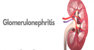
Cortical nephrocalcinosis is rare and usually occurs secondary to diffuse cortical disease injury. The calcification can be patchy or confluent. In chronic glomerulonephritis, calcium deposits are found most often in periglomerular tissue and not in the glomerulus. Nephrocalcinosis also has been reported in familial infantile nephrotic syndrome and in Alport syndrome.
Acute cortical necrosis secondary to toxemia of pregnancy, snakebite, or hemolytic-uremic syndrome can lead to patchy cortical nephrocalcinosis. Calcium deposition can start as early as 30 days after cortical necrosis. Chronic pyelonephritis and vesicoureteral reflux are also implicated. Kidney transplantation, primary hyperoxaluria, methoxyflurane abuse, autosomal recessive polycystic kidney disease, and benign nodular cortical nephrocalcinosis may be involved in cortical nephrocalcinosis, albeit rarely.
Medullary nephrocalcinosis assumes the form of small nodules of calcification clustered in each pyramid. Diagnosing the underlying renal disease on the basis of the appearance is difficult. Characteristic exceptions include papillary necrosis due to analgesic abuse and medullary sponge kidneys. In papillary necrosis, the entire papilla may be calcified, whereas in medullary sponge kidney, there is a characteristic band of calcification in the renal pyramids.
It has been suggested that when hypercalcemia is the most important factor, the first foci of calcification develop in the renal tubular cells, whereas when hypercalciuria is the major factor, the initial foci form in the interstitium.
Intraluminal tubular calcium crystals are believed to serve as potential nidi for further buildup of calcium and other stone-forming substances, including oxalate and uric acid. Whether further growth of nephroliths occurs probably depends on a number of additional factors, such as abnormal urine composition, urine flow and volume, and the presence or absence of endogenous inhibitors of crystalline formation in the urine.
Causes
Any disorder that leads to high levels of calcium in the blood or urine may lead to nephrocalcinosis. In this disorder, calcium deposits in the kidney tissue itself. Most of the time, both kidneys are affected.
Nephrocalcinosis is related to, but not the same as, kidney stones (nephrolithiasis).
Conditions that can cause nephrocalcinosis include:
- Alport syndrome
- Bartter syndrome
- Chronic glomerulonephritis
- Familial hypomagnesemia
- Medullary sponge kidney
- Primary hyperoxaluria
- Renal transplant rejection
- Renal tubular acidosis (RTA)
- Renal cortical necrosis
Other possible causes of nephrocalcinosis include:
- Ethylene glycol toxicity
- Hypercalcemia (excess calcium in the blood) due to hyperparathyroidism
- Use of certain medicines, such as acetazolamide, amphotericin B, and triamterene
- Sarcoidosis
- Tuberculosis of the kidney and infections related to AIDS
- Vitamin D toxicity
What are the Risk Factors for Nephrocalcinosis? (Predisposing Factors)
The risk factors for Nephrocalcinosis may include:
- Premature birth
- Presence of kidney stones
- Chronic kidney failure
- Sarcoidosis
- Infections
- Overuse of vitamin D supplements
- Osteoporosis
- Certain inherited conditions (such as multiple endocrine neoplasm type 1 and Bartter syndrome)
It is important to note that having a risk factor does not mean that one will get the condition. A risk factor increases one’s chances of getting a condition compared to an individual without the risk factors. Some risk factors are more important than others.
Also, not having a risk factor does not mean that an individual will not get the condition. It is always important to discuss the effect of risk factors with your healthcare provider.
What are the symptoms of nephrocalcinosis?
In many cases nephrocalcinosis doesn’t cause any early symptoms; in other cases, it is detected due to that is causing the disorder. However, people who, in addition to nephrocalcinosis, suffer from symptoms of an underlying condition kidney stones may have:
- Blood in the urine
- Fever and chills
- Nausea and vomiting
- Abdominal pain
- Pain in both sides of the back, in the groin or testicles
- Pain in the groin or testicles
In addition, late symptoms of nephrocalcinosis may be associated with chronic renal failure.
Possible Complications
Complications may include:
- Acute kidney failure
- Long-term (chronic) kidney failure
- Kidney stones
- Obstructive uropathy (acute or chronic, unilateral or bilateral)
Nephrocalcinosis diagnosis
Nephrocalcinosis may be discovered when symptoms of renal insufficiency, kidney failure, obstructive uropathy, or urinary tract stones develop.
Imaging tests can help diagnose this condition. Tests that may be done include:
- Abdominal CT scan
- Ultrasound of the kidney
Other tests that may be done to diagnose and determine the severity of associated disorders include:
- Blood tests to check levels of calcium, phosphate, uric acid, and parathyroid hormone
- Urinalysis to see crystals and check for red blood cells
- 24-hour urine collection to measure acidity and levels of calcium, sodium, uric acid, oxalate, and citrate
Nephrocalcinosis treatment
The goal of treatment is to reduce symptoms and prevent more calcium from building up in the kidneys.
Treatment will involve methods to reduce abnormal levels of calcium, phosphate, and oxalate in the blood and urine. Options include making changes in your diet and taking medicines and supplements.
Pharmacologic and other nonsurgical treatments for hypercalcemia and nephrocalcinosis include the following:
- Adequate hydration with an isotonic sodium chloride solution (the single most effective measure for reversing hypercalcemia and protecting the kidneys)
- Cinacalcet (for correction of hyperparathyroidism)
- Chemotherapeutic agents (for osteolytic malignancies)
- Steroids (to decrease intestinal calcium absorption and vitamin-D activity)
- Hydroxychloroquine (for sarcoid granulomas)
- Calcitonin or bisphosphonates (to inhibit bone resorption)
Pharmacologic and other nonsurgical treatments for macroscopic nephrocalcinosis include the following:
- Thiazide diuretics (eg, hydrochlorothiazide)
- Dietary salt restriction
- Potassium and magnesium supplementation
- Citrate supplementation (preferably as potassium citrate)
- High-dose pyridoxine
Surgery options that may be considered for urinary stones causing obstruction include the following:
- Percutaneous nephrolithotomy
- Laser and shock wave lithotripsy
- Stent placement
- Open surgery (rarely necessary)
If you take medicine that causes calcium loss, your health care provider will tell you to stop taking it. Never stop taking any medicine before talking to your provider.
Other symptoms, including kidney stones, should be treated as appropriate.
How can Nephrocalcinosis be prevented?
- Nephrocalcinosis may be prevented by seeking treatment for pre-existing disorders that may cause the condition.
- In addition, an early diagnosis and prompt treatment may reverse calcium deposits in the kidney and prevent kidney damage
- Keeping oneself hydrated by drinking plenty of fluids may also help decrease stone formation in the urinary tract
- Active research is currently being performed to explore the possibilities for treatment and prevention of disorders such as Nephrocalcinosis
- Regular medical screening at periodic intervals with tests and physical examinations are recommended