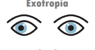
Definition of Anisometropia
Anisometropia: The condition in which the two eyes have unequal refractive power. One eye may be myopic (nearsighted) and the other hyperopic (farsighted) or one eye may be markedly stronger than the other.
Anisometropia is a serious concern in newborns and young children because it can lead to amblyopia (impaired vision in one eye). With a major degree of anisometropia, the brain cannot reconcile the difference in images coming from the two eyes. It develops a preference for the image coming from one eye and suppresses the image from the other eye and, in time, the brain loses the ability to “see” the image from the suppressed eye.
Anisometropia is derived from Greek and made up of four parts: an-, -iso-, -metr-, and -opia. Their meaning (in parentheses) is simple. An- (not) + -iso- (equal) + -metr- (measure) + -opia (vision). Literally, the measure of vision is not equal. The refractive power of the two eyes is different. The opposite of anisotropia is isometropia. In isometropia, the refractive power is equal in both eyes.
Types of Anisometropia
There are two broad categories of anisometropia: absolute anisometropia and relative anisometropia. Absolute anisometropia is then divided further.
Relative anisometropia is when the total refraction of the two eyes is the same (or at least similar enough not to qualify as anisometropic), but there is a difference in axial length between the eyes. Put in simpler terms, it is when the eyes are similar in refractive power but of different sizes. This means both eyes put out a clear image, but the images are of different sizes.
Absolute anisometropia occurs when the total refraction of the two eyes differs enough to be a problem. This condition is then further categorized, depending on how each eye refracts.
- Simple anisometropia: One eye sees normally, while the other is myopic or hypermetropic.
- Compound anisometropia: Both eyes are myopic or hypermetropic (also called ametropic).
- Mixed anisometropia: One eye is myopic; the other is hypermetropic.
- Compound astigmatic anisometropia: Both eyes are astigmatic, but to an unequal degree.
- Simple astigmatic anisometropia: One eye is either myopic or hypermetropic, and the other is astigmatic.
Causes of Anisometropia
Anisometropia may be congenital or acquired.
Congenital and developmental anisometropia: This is produced due to differential growth of each eyeball. It is hereditary in origin.
Acquired anisometropia: This is produced by
- Post cataract surgery uniocular aphakia
- Incorrect power of intraocular lens implant in patients of pseudophakia
- Eye injury
- Inadvertent surgical treatment of refractive error
- Keratoplasty in one eye
What are the risk factors for the Condition?
Medical experts still don’t know the factors that cause anisometropia, but a large difference in the refractive powers in both eyes is definitely a contributing factor.
Anisometropia symptoms
Anisometropia has no symptoms. Anisometropia is commonly found during a routine vision exam.
There are a number of potential symptoms, including:
Amblyopia (lazy eye). Usually, this is when reduced refractive power in one eye causes a lack of visual stimulation that results in insufficient information being transmitted through the optic nerve to the brain
Strabismus (crossed eyes). When a patient is unable to align both eyes. This lack of coordination prevents both eyes being able to focus on the same point in space
Diplopia (often known as double vision). The result includes:
- Eyestrain
- Headaches
- Nausea
- Light sensitivity
- Tiredness
- Dizziness
What are the complications in the case of Anisometropia?
- Inability to read, drive, play sports, or carry out routine duties due to vision issues
- Strabismus development
- Reduced quality of life brought on by the loss of stereoscopic vision, which may also raise the chance of having a car accident or having an industrial accident.
Diagnosis
If a person has anisometropia it is usually detected, or diagnosed, when the child has a vision exam. If it happens in an older person, they may notice that their vision is blurry and go in for an exam for a diagnosis. During the eye exam the doctor will check to see how well they see things when covering first one eye, then the other. It can also be diagnosed if it causes other conditions such as eyestrain or double vision.
Lab Tests and Procedures
The following lab tests and procedures are used to detect Anisometropia:
- Retinoscopy: To obtain objective measurement of refractive error of eyes
- FRIEND test: To detect the type of disease using Snellen distance vision chart
- Worth’s four dot test: To detect the type of disease using Snellen distance vision chart
Treatment for Anisometropia
Anisometropia can be treated in a number of ways. Corrective lenses are commonly used, but they only work for those with a difference of less than 4D between their eyes.
With children under the age of 12, it generally recommended that sight be corrected with contact lenses. This ensures the best possible vision in both eyes, as contact lenses can aid their development.
Older children and adults can generally use either contacts or glasses to correct their vision. Those with severe anisometropia are generally advised to use contact lenses.
The preferred method of treatment for patients with anisometropia is corrective surgery, which can sometimes permanently solve most or all of the problem. Typical surgical therapies include:
- Refractive corneal surgery. Used to improve the cornea’s refraction of light, this surgery can correct unilateral myopia, hypermetropia, and astigmatism.
- Removal of the crystalline lens. The crystalline lens is your eye’s natural lens. For some people, removing the lens can actually improve their overall vision. The sight in that eye can then be adjusted with special glasses or further surgeries.
- Intraocular lens implantation. Intraocular lens implantation (IOL) is a relatively common surgery for those with a cataract or astigmatism in their eye that is seriously impacting their life. While there are several variations of this surgery, the eye is generally cut precisely so a doctor can break up the natural lens. Then, a plastic lens is put in its place to correct vision.
- Phakic IOL. This intraocular lens implantation is similar to the above surgery, but it does not remove the crystalline lens. Instead, the intraocular lens is placed in the eye with the natural lens. The two will then work together to improve vision.
Any surgery carries risks. While the above surgeries are not especially dangerous, complications are still possible that could lead to blindness, a reduction in visual clarity, or infection. More commonly, you may experience redness or swelling.
Talk to your doctor about any concerns you have. Report any problems you experience to your doctor immediately.
Follow your doctor’s recovery plan closely. During this vulnerable recovery time, it’s easy to damage the eye. Ask your doctor what to expect, what behaviors to avoid, and roughly how long it will take to recover.
Do not touch your eye as it heals, even though it may itch or ooze fluid. Your doctor will likely prescribe medicated eye drops to aid in your recovery.
How can I prevent Anisometropia?
There’s currently no way to prevent refractive errors.