Definition
Bones naturally grow and change as your child gets older. However, throughout a long process, abnormalities can sometimes occur. An aneurysmal bone cyst (which is often abbreviated ABC) is one such abnormality. ABCs are blood-filled, fibrous cysts that expand the bone and can cause pain, swelling, and fractures. They are benign cysts (non-cancerous) that don’t spread.
Aneurysmal Bone Cyst
ABCs are most common during a child’s teenage years. They can occur in virtually any bone in the arms, legs, trunk or skull as well as the vertebrae and knee. While it’s benign, it can be quite destructive, because it deforms the bone and can cause fractures. ABC’s don’t spread but can be quite damaging to the bone and can come back after they are removed. With surgery, these cysts are highly curable, although they do grow back in some cases.
Types of Aneurysmal Bone Cyst
An aneurysmal bone cyst usually falls into one of two categories: active and aggressive.
- An active ABC is one that could deform the bone it’s growing in, but remains contained in the bone.
- While an aggressive ABC extends beyond the bone to the nearby soft connective tissues. Rarely, will aggresive cysts go away without treatment.
Both types can cause pain and swelling and, in rare cases, fractures in the involved bone. Aneurysmal bone cysts do not generally go away on their own.
Pathophysiology
Aneurysmal bone cysts consist of blood-filled spaces of variable size that are separated by connective tissue containing trabeculae of bone or osteoid tissue and osteoclast giant cells. They are not lined by endothelium. A fine-needle aspiration cytology is usually nondiagnostic, often dominated by fresh blood.
Although often primary, up to a third of aneurysmal bone cysts are secondary to an underlying lesion (e.g. fibrous dysplasia, osteosarcoma, giant cell tumor, chondromyxoid fibroma, non-ossifying fibroma, chondroblastoma).
A variant of aneurysmal bone cysts is the giant cell reparative granuloma which is usually seen in the tubular bones of the hands and feet as well as in the craniofacial skeleton. Occasionally they are also seen in appendicular long bones where they are known as solid aneurysmal bone cysts. Histologically these two entities are identical.
Aneurysmal Bone Cyst Risk factors
No clear risk factors have been established for the development of Aneurysmal Bone Cyst. However, the following are thought to be associated with the condition:
- Trauma
- Preexisting bone lesions, such as:
- Giant cell tumor
- Chondroblastoma
- Fibrous dysplasia
It is important to note that having a risk factor does not mean that one will get the condition. A risk factor increases ones chances of getting a condition compared to an individual without the risk factors. Some risk factors are more important than others.
Also, not having a risk factor does not mean that an individual will not get the condition. It is always important to discuss the effect of risk factors with your healthcare provider.
Causes of Aneurysmal Bone Cyst
- The cause of these cysts is unknown and controversial. They’re believed to grow in response to a disturbance of the blood vessels in the involved bone. They may grow because of a pre-existing tumor. In half of all cases, a pre-existing tumor, such as fibrous dysplasia, nonossifying fibroma, a solitary bone cyst or osteosarcoma also exists.
- Abnormalities in the chromosomes (karyotype) of the tumor cells have been described, but the significance of these findings is unclear.
- There is no definitive explanation for why these cysts occur. It’s important to know that there’s nothing that you could have done (or not done) that would have prevented your child’s cyst from developing.
Symptoms
While symptoms may vary child-to-child, the most common include:
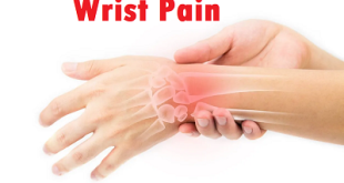
- Swelling
- Mild to severe neurological problems (if the cyst is in your child’s spine)
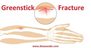
- A fracture caused by the cyst (rarely)
Swelling in bone
It’s important to understand that the symptoms of the aneurysmal bone cyst may resemble other medical problems, some of them which are very common and easy to treat, others which could be more serious.
Your child may experience symptoms differently. Therefore, it is important to be evaluated by a physician to get an accurate diagnosis. Always consult your child’s physician if you have concerns.
Complications
Recurrence
Younger age and open physes are known risk factors for ABC recurrence. Histologically, an ABC with a higher proportion of cellular tissue relative to the amount of osteoid and fibrillary content is a prognostic indicator of recurrence. In addition, the presence of higher mitotic indices is a risk factor for an increased recurrence rate.
Malignant Transformation
Malignant transformation is rare and can occur with and without the use of adjuvants during primary treatment. Malignant transformation from ABC to fibroblastic osteosarcoma and telangiectatic osteosarcoma has been reported.
Diagnosis and test
If an aneurysmal bone cyst is not treated it can cause pain, fractures, disrupt growth and cause neurological symptoms.
Clinical experts use a variety of diagnostic tests to diagnose aneurysmal bone cysts, including:
- X-rays, which produce images of bones.
- Magnetic resonance imaging (MRI), which uses a combination of large magnets, radiofrequencies and a computer to produce detailed images of organs, soft tissues, muscles, ligaments and other structures within the body. Your child is exposed to no radiation during an MRI.
- Computed tomography (CT) scan, which uses a combination of X-rays and computer technology to examine bones and produces cross-sectional images (“slices”) of the body.
- EOS imaging, an imaging technology that creates 3-dimensional models from two flat images. Unlike a CT scan, EOS images are taken while the child is in an upright or standing position, enabling improved diagnosis due to weight-bearing positioning.
- Angiography, a radiograph-type X-ray test which reveals the inside of blood vessels and organs.
- Needle biopsy, which is a procedure where a doctor places a small needle through the skin and into the lesion to withdraw a small sample of the abnormal tissue. The tissue is analyzed to confirm any findings.
In addition to diagnosing the specific type of growth your child may have, these tests will also help determine the size and location of the tumor. All of this information is crucial in determining the best treatment options for your child.
Treatment and medications
Sclerotherapy
- “Washing” the ABC with medicine that causes it to fill in with bone. This is done by placing needles in the ABC under imaging guidance.
- Serial treatments are performed.
- Treating aneurysmal bone cysts by removing fluid from the cyst and replacing it with medicine called Doxycyline
Sclerotherapy
Embolization
Blocking the arterial blood supply using tiny particles or glue.
This is often done in preparation for surgical resection/removal.
The procedure typically requires 30 – 60 minutes to complete.
Typically, sedation or anesthesia will be used to help facilitate the successful completion of the procedure. This helps to minimize anxiety for the child and decrease risk of complications as a result of movement during the procedure.
- The child will be positioned on the imaging table.
- The imaging technique used is a mobile X ray tube (fluoroscopy) that can also make a CT image used for needle guidance.
- The skin surface will be cleaned to allow for a sterile skin puncture site.
- Local anesthetic will be introduced to help with comfort at the expected skin puncture site prior to a small skin nick being made to permit insertion of the needle into the ABC.
- Contast is injected to fill the ABC and determine volume and blood flow to the ABC.
- Medicine is introduced to scar the inside of the ABC which initiates the healing process.
Typically patients go home the same day of the procedure with pain medicine. Your child may have some soreness at the site of treatment. Typically this can be well controlled with ibuprofen or acetaminophen (Tylenol®) or stronger medications.
Prevention of Aneurysmal Bone Cyst
- Current medical research has not established a way of preventing Aneurysmal Bone Cyst
- Regular medical screening at periodic intervals with blood tests, scans, and physical examinations, are mandatory for those who have already endured the tumor, due to its metastasizing potential and possibility of recurrence. Often several years of active vigilance is necessary


what if the bone ache without swelling,what to do?
It might be a symptom of Paget’s disease. So it would be better to consult a doctor to diagnose the problem.