Definition
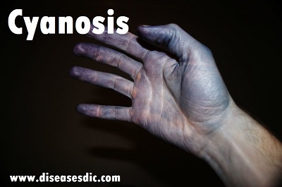
Cyanosis is the medical term for a bluish color of the skin and the mucous membranes due to an insufficient level of oxygen in the blood. For example, the lips and fingernails may show cyanosis. Cyanosis can be evident at birth due to the presence of a heart malformation that permits blood that is not fully oxygenated to enter the arterial circulation. Cyanosis can also appear at any time later in life and often accompanies conditions in which lung function is compromised (resulting in an inability to fully oxygenate the blood) or conditions in which the heart’s pumping function is compromised.
The presence of abnormal forms of hemoglobin or other abnormalities of the blood cells can also sometimes cause cyanosis. The medical term for lowered oxygen levels is hypoxia; the term anoxia refers to the absence of oxygen. Pseudocyanosis is the appearance of cyanosis that is not associated with reduced oxygen delivery to tissues. Most causes are related to the ingestion of metals (such as silver or lead) or drugs/toxins.
History of Cyanosis
The name cyanosis literally means the blue disease or the blue condition. It is derived from the color cyan, which comes from cyanós (κυανός), the Greek word for blue. It is postulated by Dr. Christen Lundsgaard that cyanosis was first described in 1749 by Jean-Baptiste de Sénac, a French physician who served King Louis XV. De Sénac concluded from an autopsy that cyanosis was caused by a heart defect that led to the mixture of arterial and venous blood circulation. But it was not until 1919 when Dr. Lundsgaard was able to derive the concentration of deoxyhemoglobin (8 volumes percent) that could cause cyanosis.
Pathophysiology
Cyanosis typically occurs when the amount of oxygen bound to hemoglobin is very low. Oxygen in the blood is carried in two physical states. Approximately 2% is dissolved in plasma and the other 98% is bound to hemoglobin. The presence of cyanosis might be an indication of inadequate oxygen delivery to the peripheral tissues. It also could be related to increased oxygen extraction by the peripheral tissues. Several factors play a significant role in oxygen delivery to the end organs. Oxygen delivery is the product of the cardiac output and arterial oxygen content. Cardiac output is determined by the preload, afterload, and contractility. The arterial oxygen content is the sum of oxygen bonded to hemoglobin and dissolved in plasma, approximately 1.34 mL per 1 g of hemoglobin and 0.003 mL of oxygen per 100 mL of plasma.
Typically, when the level of deoxygenated hemoglobin is around 3 to 5 g/dL, cyanosis becomes very evident. The presence of jaundice, skin color, ambient temperature, or light exposure might affect the assessment of cyanosis. Anemia or polycythemia also plays a role in cyanosis. The level of hypoxia required to produce clinically evidenced cyanosis varies for a given level of hemoglobin. Cyanosis is more difficult to discern when the level of hemoglobin is low. In other words, cyanosis might not be clinically evident in a patient with severe anemia.
Types of Cyanosis
Cyanosis is of two types – central and peripheral. The characteristic features of these two types of cyanosis include the following:
Central Cyanosis
This condition occurs due to heart and lung conditions, as well as the occurrence of abnormal forms of hemoglobin, such as methemoglobin and sulfhemoglobin in the blood. These are responsible for the blue-purple discoloration of the tongue and the lining of the oral cavity. It has been observed that central cyanosis can be accompanied by peripheral cyanosis. However, in the absence of peripheral cyanosis, the toes and fingers usually exhibit a warm sensation to touch.
Peripheral Cyanosis
This occurs due to the decreased blood flow to the peripheral parts (extremities) of the body, such as the fingers, toes and especially the nail beds. Peripheral cyanosis is accentuated by the stagnation of arterial blood in the capillaries of the periphery and release of most of the oxygen to the tissues. As a result, the level of deoxygenated blood increases in the capillaries and veins. This can occur due to congestive heart failure and shock, both of which exhibit sluggish blood circulation and rapid fall in blood pressure in the capillaries and veins. Peripheral cyanosis is also precipitated by exposure to extremely cold temperatures such as higher altitudes, as well as vascular diseases. It responds well to warming-up the limbs.
Peripheral Cyanosis
Risk factors
There are a few factors that can increase the risk of a person developing Cyanosis. Some of the Cyanosis risk factors are written as follows:
- History of any congenital heart disease
- People who go to high altitudes for the first time
- Exposure to Carbon Monoxide
- Any kind of seizure
- History of lungs or alveoli diseases like Asthma, Chronic bronchitis, or Bronchiectasis
- Living in very cold conditions
Causes of Cyanosis
Oxygen is what makes blood red. Getting enough oxygen through your lungs and circulating it effectively throughout your body is what gives your skin a normal pink or red tinge (regardless of your skin tone).
Blood that doesn’t have much oxygen in it is carrying mainly waste carbon dioxide from your cells to be exhaled from your lungs. This oxygen-poor blood is darker in color and more bluish-red than true red.
It’s normal for your veins to show this bluish color since veins deliver blood with its waste cargo back to the heart and lungs to get rid of the carbon dioxide.
But when parts of your body turn blue or purple due to cyanosis, there’s an underlying issue that’s limiting blood flow or oxygen that must be addressed immediately.
Cyanosis can be caused by a wide variety of medical conditions, such as:
- Chronic obstructive pulmonary disease (COPD)
- Pulmonary hypertension (a complication of COPD)
- Pneumonia
- Infections of the respiratory tract
- Asthma
- Congestive heart failure
- Raynaud’s phenomenon, a condition that causes your blood vessels to narrow, mainly in your fingers and toes
- Epiglottitis is a serious condition involving swelling of the small flap in your throat that covers your windpipe
- Hypothermia
- Seizures
- Drug overdose
- Suffocation
Symptoms
Some heart defects cause major problems right after birth.
The main symptom of cyanosis is a bluish color of the lips, fingers, and toes that is caused by the low oxygen content in the blood. It may occur while the child is resting or only when the child is active.
Some children have breathing problems (dyspnea). They may get into a squatting position after physical activity to relieve breathlessness.
Others have spells, in which their bodies are suddenly starved of oxygen. During these spells, symptoms may include:
- Anxiety
- Breathing too quickly (hyperventilation)
- A sudden increase in the bluish color of the skin
Infants may get tired or sweat while feeding and may not gain as much weight as they should.
Fainting (syncope) and chest pain may occur.
Other symptoms depend on the type of cyanotic heart disease, and may include:
- Feeding problems or reduced appetite, leading to poor growth
- Grayish skin
- Puffy eyes or face
- Tiredness all the time
A baby with Cyanosis
Complications of Cyanosis
Because cyanosis can be due to serious diseases, failure to seek treatment can result in serious complications and permanent damage. Once the underlying cause is diagnosed, it is important for you to follow the treatment plan that you and your healthcare professional design specifically for you to reduce the risk of potential complications including:
- Gangrene or ulceration
- Heart failure
- Loss of limb
- Respiratory failure
- Sepsis (life-threatening inflammatory reaction to infection)
Diagnosis and test
Bluish skin is usually a sign of something serious. If the normal color does not return when your skin is rubbed or warmed, it is important to get medical attention right away to determine the cause.
The physical examination performed by your doctor will include listening to your heart and lungs. You may also have to undergo a series of other clinical tests.
Apart from the clinical assessment of hypoxemia, the diagnosis of Cyanosis may also include the following investigations:
- Arterial Blood Gas test: Measures the acidity and levels of carbon dioxide and oxygen in your blood.
- Complete Blood Count: Haemoglobin levels are increased with the prevalence of chronic Cyanosis. The white cell count increases in conditions like pneumonia and pulmonary embolism.
- ECG: Taken to completely rule out the prevalence of cardiac abnormalities.
- Chest X-ray: This is taken to rule out conditions like pneumonia, pulmonary infarction, and cardiac failure.
- Ventilation-perfusion scan or Pulmonary Angiography is taken to rule out pulmonary causes
- Echocardiography will serve to look for the presence of any cardiac defects.
- Hemoglobin spectroscopy will look for methemoglobinemia or sulfhemoglobinemia.
- Digital Subtraction Angiography: is done to completely rule out conditions like acute arterial occlusion.
- A duplex Doppler or Venography can detect the prevalence of acute venous occlusion.
Treatment and medications
Treating cyanosis completely depends on treating the underlying cause. Timely treatment can help to prevent further complications of low blood oxygen.
- Heat therapy is an application of mild heat to the affected areas that can improve the symptoms of peripheral cyanosis.
- In children, the inability to feed results in secondary cyanotic heart disease, which leads to metabolic abnormalities such as hypocalcemia and hypoglycemia. Thus metabolic abnormalities need to be corrected.
- Oxygen therapy is provided initially to reverse the hypoxia condition (low level of oxygen). The goal is to restore oxygenated blood supply as early as possible.
- Surgical intervention is indicated in babies with congenital heart defects such as Fallot’s tetralogy. Open heart surgery is indicated immediately soon after birth to correct the defects. For less severe defects, surgery is carried out when the baby reaches around three to six months of age.
- Treatment for peripheral cyanosis relaxes the blood vessels and may include antidepressants, drugs used for treating erectile dysfunction (PDE5 Inhibitors), and antihypertensive medications – ACE inhibitors (angiotensin-converting enzyme inhibitors), and Diuretics (increases the excretion of water through kidneys) are prescribed.
- In the case of Raynaud’s phenomenon, lifestyle modifications are part of treatment which includes limits or avoiding things that can constrict blood vessels, such as
- Intake of caffeine and nicotine slowdowns the blood flow and narrows blood vessels. Thus the limited intake of both nicotine and caffeine is beneficial.
- Certain medications, like decongestants, migraine medications and birth control pills, beta-blockers should be avoided, which further cause constriction of blood vessels and restricts blood flow.
Other lifestyle modifications include exercising regularly, keeping extremities warm, reducing stress, and avoiding rapid temperature changes.
Prevention of Cyanosis
Cyanosis can prove fatal if not treated on time. Hence, it is better to have Cyanosis prevention rather than taking any risk. A few preventive methods for this condition are as follows:
- Try to avoid edible or smoking products that contain caffeine and nicotine. These substances constrict your blood vessels and increase the chance of Cyanosis development.
- Don’t go to high altitudes if you have developed heart conditions.
- If you go to some cold area, make sure you have medical help in your reach.
- Take oxygen cylinders with you while going to high-altitude places like mountains.

