Definition
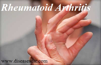
Rheumatoid arthritis (RA) is an autoimmune disease in which the body’s immune system – which normally protects its health by attacking foreign substances like bacteria and viruses – mistakenly attacks the joints. This creates inflammation that causes the tissue that lines the inside of joints (the synovium) to thicken, resulting in swelling and pain in and around the joints. The synovium makes a fluid that lubricates joints and helps them move smoothly.
Rheumatoid arthritis
If inflammation goes unchecked, it can damage cartilage, the elastic tissue that covers the ends of bones in a joint, as well as the bones themselves. Over time, there is loss of cartilage, and the joint spacing between bones can become smaller. Joints can become loose, unstable, painful and lose their mobility.
History
Arthritis and diseases of the joints have been plaguing mankind since ancient times. In around 1500 BC the Ebers Papyrus described a condition that is similar to rheumatoid arthritis. This is probably the first reference to this disease.
There is evidence of rheumatoid arthritis in the Egyptian mummies as found in several studies. G. Elliot in his studies found that rheumatoid arthritis was a prevalent disease among Egyptians.
In the Indian literature, Charak Samhita (written in around 300 – 200 BC) also described a condition that describes pain, joint swelling and loss of joint mobility and function. Hippocrates described arthritis in general in 400 BC. He however did not describe specific types of arthritis. Galen between 129 and 216 AD introduced the term rheumatismus.
Paracelsus (1493-1511) suggested that substances that could not be passed in urine got stored and collected in the body especially in the joints and this caused arthritis. Ayurveda in ancient Indian medicine also considered arthritis as one of the Vata. Practitioners attributed rheumatic disorders to humors (rheuma).
Thomas Sydenham first described a disabling form of chronic arthritis that was described later by Beauvais in 1880. Brodie went on to show the progressive nature of this disease and found how rheumatoid arthritis affected the tendon sheaths and sacs of synovium in the joints. He found how there was synovial inflammation or synovitis and cartilage damage associated with rheumatoid arthritis.
Epidemiology
Worldwide, the annual incidence of RA is approximately 3 cases per 10,000 population, and the prevalence rate is approximately 1%, increasing with age and peaking between the ages of 35 and 50 years. RA affects all populations, though it is much more prevalent in some groups (eg, 5-6% in some Native American groups) and much less prevalent in others (eg, black persons from the Caribbean region).
First-degree relatives of individuals with RA are at 2- to 3-fold higher risk for the disease. Disease concordance in monozygotic twins is approximately 15-20%, suggesting that nongenetic factors play an important role. Because the worldwide frequency of RA is relatively constant, a ubiquitous infectious agent has been postulated to play an etiologic role.
Women are affected by RA approximately 3 times more often than men are, but sex differences diminish in older age groups. In investigating whether the higher rate of RA among women could be linked to certain reproductive risk factors, a study from Denmark found that the rate of RA was higher in women who had given birth to just 1 child than in women who had delivered 2 or 3 offspring. However, the rate was not increased in women who were nulliparous or who had a history of lost pregnancies. Time elapsed since pregnancy is also significant. In the 1- to 5-year postpartum period, a decreased risk for RA has been recognized, even in those with higher-risk HLA markers.
Types of rheumatoid arthritis
As of now, there is a primary way of defining the type of rheumatoid arthritis a patient has. First of all, doctors determine whether the patient has seropositive rheumatoid arthritis or seronegative rheumatoid arthritis. This difference can determine treatment options.
Seropositive
- Rheumatoid arthritis patients who are classified as seropositive have the presence of anti-cyclic citrullinated peptides (anti-CCPs) in their blood test results. These are also referred to as anti-citrullinated protein antibodies (ACPAs). These are the antibodies that attack the body and produce the symptoms of rheumatoid arthritis.
- Between 60 and 80 % of rheumatoid arthritis patients test positive for the presence of anti-CCPs, meaning it is a reliable indicator for diagnosis. The presence of these antibodies can be detected as early as 5 to 10 years before clinical rheumatoid arthritis symptoms appear.
Seronegative
- It’s still possible for patients to develop rheumatoid arthritis without the presence of antibodies in their blood. This is referred to as seronegative type rheumatoid arthritis. Seronegative patients are those who do not test positive for the anti-CCPs or another antibody called rheumatoid factor.
- Though seronegative patients lack the antibodies that help doctors diagnose the condition, they can still be diagnosed with rheumatoid arthritis in a number of ways. These include the demonstration of clinical rheumatoid arthritis symptoms, as well as X-ray results indicating patterns of cartilage and bone deterioration.
- Though it’s possible for seronegative patients to have milder rheumatoid arthritis symptoms than seropositive patients, this isn’t always the case. It can still depend on a number of factors, including genetics and other underlying conditions as well.
- Unfortunately, many seronegative patients may not respond to typical rheumatoid arthritis symptoms. This provides further motivation for researchers to identify rheumatoid arthritis sub-types in order to provide treatment for those who don’t have any long-term solutions as of now.
Risk factors of rheumatoid arthritis
Factors that may increase your risk of rheumatoid arthritis include:
Your sex: Women are more likely than men to develop rheumatoid arthritis.
Age: Rheumatoid arthritis can occur at any age, but it most commonly begins between the ages of 40 and 60.
Family history: If a member of your family has rheumatoid arthritis, you may have an increased risk of the disease.
Smoking: Cigarette smoking increases your risk of developing rheumatoid arthritis, particularly if you have a genetic predisposition for developing the disease. Smoking also appears to be associated with greater disease severity.
Environmental exposures: Although uncertain and poorly understood, some exposures such as asbestos or silica may increase the risk for developing rheumatoid arthritis. Emergency workers exposed to dust from the collapse of the World Trade Center are at higher risk of autoimmune diseases such as rheumatoid arthritis.
Obesity: People who are overweight or obese appear to be at somewhat higher risk of developing rheumatoid arthritis, especially in women diagnosed with the disease when they were 55 or younger.
Causes
- Rheumatoid arthritis occurs when your immune system attacks the synovium the lining of the membranes that surround your joints.
- The resulting inflammation thickens the synovium, which can eventually destroy the cartilage and bone within the joint.
- The tendons and ligaments that hold the joint together weaken and stretch. Gradually, the joint loses its shape and alignment.
- Doctors don’t know what starts this process, although a genetic component appears likely. While your genes don’t actually cause rheumatoid arthritis, they can make you more susceptible to environmental factors such as infection with certain viruses and bacteria that may trigger the disease.
Symptoms of rheumatoid arthritis
While early symptoms of rheumatoid arthritis can actually be mimicked by other diseases, the symptoms are very characteristic of rheumatoid disease. Rheumatoid arthritis symptoms and signs include the following:
- Fatigue
- Joint pain
- Joint tenderness
- Joint swelling
- Joint redness
- Joint warmth
- Joint stiffness
- Loss of joint range of motion
- Limping
- Joint deformity
- Many joints affected (polyarthritis)
- Both sides of the body affected (symmetric)
- Loss of joint function
- Anemia
- Fever
Diagnosis and test
Medical History
The doctor will ask about personal and family medical history as well as recent and current symptoms (pain, tenderness, stiffness, difficulty moving).
Physical Exam
The doctor will examine each joint, looking for tenderness, swelling, warmth and painful or limited movement. The number and pattern of joints affected can also indicate RA. For example, RA tends to affect joints on both sides of the body. The physical exam may reveal other signs, such as rheumatoid nodules or a low-grade fever.
Blood Tests
The blood tests will measure inflammation levels and look for biomarkers such as antibodies (blood proteins) linked with RA.
Inflammation
Erythrocyte sedimentation rate (ESR, or “sed rate”) and C-reactive protein (CRP) level are markers of inflammation. A high ESR or CRP is not specific to RA, but when combined with other clues, such as antibodies, helps make the RA diagnosis.
Antibodies
Rheumatoid factor (RF) is an antibody found in about 80 percent of people with RA during the course of their disease. Because RF can occur in other inflammatory diseases, it’s not a sure sign of having RA. But a different antibody – anti-cyclic citrullinated peptide (anti-CCP) – occurs primarily in patients with RA. That makes a positive anti-CCP test a stronger clue to RA. But anti-CCP antibodies are found in only 60 to 70 percent of people with RA and can exist even before symptoms start.
Imaging Tests
An X-ray, ultrasound or magnetic resonance imaging scan may be done to look for joint damage, such as erosions – a loss of bone within the joint and narrowing of joint space. But if the imaging tests don’t show joint damage that doesn’t rule out RA. It may mean that the disease is in an early stage and hasn’t yet damaged the joints.
Treatment and medications
- Treatment for rheumatoid arthritis is aimed at reducing inflammation to the joints, relieving pain, minimizing any disability caused by pain, joint damage or deformity, and either slowing down or preventing damage to the joints. There is no current cure for rheumatoid arthritis.
- With the help of an occupational therapist and physical therapist (UK: physiotherapist) patients can learn how to protect their joints. Depending on the degree of damage to the joints, surgery may sometimes be needed.
- If the patient has had inflamed joints for over six weeks the GP (general practitioner, primary care physician) will most likely refer him/her to a rheumatologist (an arthritis specialist doctor), so that diagnosis can be confirmed and treatment started as soon as possible.
Medications for rheumatoid arthritis
During the initial stages of the disease the doctor will usually prescribe medications that are known to have the fewest side effects. As the disease progresses, stronger medications may be required. Many rheumatoid arthritis medications have potentially serious side effects.
1) Nonsteroidal anti-inflammatory drugs (NSAIDs)
NSAIDs are used for pain relief as well as reducing inflammation. Examples include Advil or Motrin, which are both OTC (over the counter, no prescription required). NSAIDs will not slow down the progression of the disease. When taken in high doses or over a long period they may cause complications. Side effects may include:
- A higher risk of bruising
- Gastric ulcers
- Hypertension – high blood pressure
- Kidney damage
- Liver damage
- Some heart problems
- Stomach bleeding
- Tinnitus – ringing in the ears
Cox-2 selective inhibitors, another type of NSAID, are designed to be less harmful for the stomach. However, some research has linked them to a higher risk of strokes, hypertension, heart disease and heart attacks. If the patient has a history of hypertension, high cholesterol or smokes the doctor needs to be told.
2) Corticosteroids
Corticosteroids are effective at reducing inflammation, pain, as well as slowing down joint damage. They are usually recommended when NSAIDs have not helped. If the patient has a single inflamed joint the doctor may inject the steroid into the joint. Effective relief is usually felt rapidly and the effect can last from weeks to months, depending on the severity of symptoms.
Examples include prednisone (Lodotra) and methylprednisolone (Medrol). Corticosteroids are generally used for acute symptoms (short term flare ups) – the dosage is then gradually reduced (tapered off). Long term use can have serious side effects. Side effects may include:
- A higher risk of bruising
- Cataracts
- Diabetes
- Round face
- Weight gain
- Osteoporosis
- Glaucoma
- Muscle weakness
- Thinning of the skin
3) DMARDs (disease-modifying antirheumatic drugs)
This medication may slow down the progression of the disease, as well as preventing permanent damage to the joints and other tissues. The earlier the patient starts taking a DMARD the more effective it will be.
It may take from four to six months before the patient starts noticing any beneficial effects. It is important to keep taking the medication even if initially it does not appear to be working. Some patients may have to try different types of DMARD before hitting on the most suitable one. This medication is usually taken indefinitely.
Examples include leflunomide (Arava), methotrexate (Rheumatrex, Trexall), sulfasalazine (Azulfidine), minocycline (Dynacin, Minocin), and hydroxychloroquine (Plaquenil). Side effects may include:
- Liver damage
- Bone marrow suppression
- Lung infections (severe)
4) Immunosuppressants
As rheumatoid arthritis is an auto-immune disease, suppressing the immune system helps reduce the damage to good tissue. Examples include cyclosporine (Neoral, Sandimmune, Gengraf), azathioprine (Imuran, Azasan), and cyclophosphamide (Cytoxan).
Tumor necrosis factor-alpha inhibitors (TNF-alpha inhibitors) – the human body produces tumor necrosis factor-alpha (TNF-alpha). TNF-alpha is an inflammatory substance. TNF-alpha inhibitors are used for the reduction of pain, morning stiffness and swollen or tender joints. Results are usually noticed within two weeks of starting treatment. Examples include (Enbrel), infliximab (Remicade) and adalimumab (Humira). Possible side effects include:
- A higher risk of infection
- Blood disorders
- Congestive heart failure
- Demyelinating diseases – erosion of the myelin sheath that normally protects nerve fibers, exposing the fibers, resulting in problems in nerve impulse conduction. This may affect several physical systems.
- Irritation at the injection site
- Lymphoma
Occupational therapy
An occupational therapist can help the patient learn new and effective ways of carrying out daily tasks so that stress to painful joints is minimized. For example, if the patient has sore arms and wants to push open a door, it may be better to lean into it rather than using the arms.
If the patient has painful fingers a specially devised gripping and grabbing tool may help.
Surgery
If the above-mentioned treatments have not been effective enough, the doctor may consider surgery to repair damaged joints, allowing the patient to subsequently use that joint again. Surgical intervention may also help correct deformities, or reduce pain. The following procedures may be considered:
Arthroplasty – Total replacement of the joint. The damaged parts are surgically removed and a prosthesis (artificial joint) made of metal and plastic is inserted.
Tendon repair – If the tendons around the joint are loosened or ruptured, surgery may help restore them.
Synovectomy – This involves the removal of the joint lining, if the synovium (lining around the joint) is inflamed and causing pain.
Arthrodesis – If a joint replacement is not an option, the joint may be surgically fixed to promote a bone fusion; the joint is realigned or stabilized. Also called artificial ankylosis, syndesis.
Lifestyle
When a flare-up occurs the patient should rest as much as possible. Exerting very swollen and painful joints frequently results in worsening symptoms.
Generally, when flare ups are not present, the patient should exercise regularly; this will help their general health and mobility. If rheumatoid arthritis has caused muscles around the joints to become weak, exercise will help strengthen them. Exercises that do not strain the joints are best, such as swimming. A qualified physical therapist (UK: physiotherapist) can teach the patient exercises that improve mobility.
Applying heat or cold
- Tense and painful muscles may benefit from the application of heat. A 15 minute hot bath or shower may help. Some people find that using a hot pack or an electric heating pad (set at lowest setting) helps.
- Pain may be dulled with cold treatment. The numbing effect of cold may also decrease muscle spasms. Patients with poor circulation or numbness should not use cold treatments. Examples of cold treatment include cold packs, soaking the affected joint in cold water, and ice massage.
- Some people benefit from placing the affected joints in warm water for a few minutes, followed by cool water for one minute; repeating the cycle for about 30 minutes, ending with a warm water soak.
Relaxation
Finding ways of alleviating mental stress may help control pain. Examples include hypnosis, guided imagery, deep breathing and muscle relaxation.
Complementary therapies
These are commonly used by people with rheumatoid arthritis. Few studies have been carried out on how effective they are. Examples include:
- Acupuncture
- Chiropractic
- Electrotherapy
- Hydrotherapy
- Massage
- Nutritional supplements – for example, fish oil, glucosamine sulphate and chondriotin.
- Osteopathy
Prevention of rheumatoid arthritis
- Screening and follow-up of people at risk of developing RA is appropriate for developing prevention programs for the disease. First degree relatives of RA patients, twins of RA patients, autoantibody-positive individuals, populations with high disease prevalence rate were screened in various prospective studies.
- However, these studies are expensive considering the yield as relatively low prevalence rate of RA and RA-related autoantibodies limit the statistical power of these studies.
- Large-scale screening to identify individuals at high risk of developing RA in the future (genetic factors and RA-related antibodies) will be very expensive. Low prevalence of autoantibodies in RA requires large-scale screening to identify at-risk individuals.
- As these studies are not suitable for developing countries, prevention of RA by modifying environmental factors is emerging as a cheap and effective strategy to prevent RA.
- Patients who are symptomatic definitely need treatment. Palindromic rheumatism patients often respond to antimalarials and other disease-modifying agents. Many studies show that patients with undifferentiated arthritis benefit from methotrexate.

