Definition
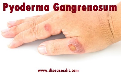
Pyoderma gangrenosum (PG) is a rare inflammatory neutrophilic dermatosis a group of conditions in which neutrophils (a type of white blood cells that protect us from infections) quickly react and infiltrate affected tissue often after some sort of abrasion or injury. PG usually begins as a small bump or sore before growing into a larger, painful open wound (skin ulcer).
Skin ulcers associated with PG have elevated borders, with the skin surrounding the wound becoming red or purple. PG ulcers are often exposed and extremely sensitive, which can lead to concerns of infection, pain management, scarring, mental issues, and potential loss of mobility.
Pyoderma gangrenosum
Epidemiology
Pyoderma gangrenosum occurs in about 1 in 100,000 persons each year in the United States. Although pyoderma gangrenosum affects both sexes, a slight female predominance may exist. All ages may be affected by the disease, but it predominantly occurs in the fourth and fifth decades of life. Children account for only 3-4% of the total number of cases. (Nothing is clinically distinctive about pyoderma gangrenosum in children and adolescents other than the age of the patients.)
Pyoderma gangrenosum pathophysiology
Much like classic PG, the most common finding was neutrophil infiltration/acute inflammatory infiltrate. Necrosis, dermal abscesses, and chronic inflammatory infiltrate were also described. Histopathologic findings depend on the site and stage of the biopsy, with early lesions or biopsies from the periphery showing moderate perivascular lymphocytic infiltrate associated with vascular endothelial swelling. More advanced lesions and central areas show abscess formation with ulceration and dense lymphocytic infiltration around (and involving) blood vessels, with thrombosis and extravasation of erythrocytes. In the late stages, the disease is characterized by almost complete replacement of the dermis by a neutrophil-laden inflammatory cell infiltrate, in which only the main cutaneous adnexal structures (such as hair follicles) remain intact. Two thirds of patients with ulcerative PG have associated systemic diseases, particularly arthritis, inflammatory bowel disease (IBD), monoclonal gammopathy, or malignancy.
Types of Pyoderma gangrenosum
There are four recognized clinical variants of PG classical or ulcerative, pustular, bullous, and vegetative or granulomatous. Some patients develop only a particular subtype, whereas others may have more than one subtype simultaneously. Some subtypes show preferential associations with underlying diseases, an eg, pustular variant in patients with IBD.
Classical PG, also known as ulcerative type, is characterized by rapidly progressive painful ulcers, which typically have undermined, overhanging, dusky purple edges, with surrounding induration and erythema. The base of the ulcer commonly has granulation tissue, and occasionally, necrotic tissue and purulent exudate. Classic PG ulcers are aseptic, although superinfection may occur.
Pustular PG variant is characterized by multiple sterile pustules with a surrounding erythematous halo, generally arising on the trunk and extensor aspect of the limbs. Histopathology reveals a dermal neutrophilic infiltrate and subcorneal neutrophilic micropustules. This variant is commonly associated with IBD, and has a tendency to remit with IBD control.
Bullous PG typically presents with grouped vesicles that rapidly spread and coalesce to form large bullae, and further develop into ulcerations, showing central necrosis and a peripheral halo of erythema. This variant generally occurs at atypical sites, on the dorsal surface of the hands, extensor aspects of the arms, or on the head, and is mostly seen in patients with lymphoproliferative diseases, as a paraneoplastic phenomenon. In patients with hematologic disease, the presence of PG may signify malignant transformation, and is suggestive of poor prognosis or aggressive disease.
Vegetative or superficial granulomatous PG is characterized by a solitary, erythematous, ulcerated plaque, lacking the violaceous border that is typically present in the classical variant. Histologically, it is characterized by granulomas with a three-layered structure: the center contains the neutrophilic inflammation, surrounded by a palisade of histiocytes, which is in turn rimmed by a lymphocytic infiltrate. This is the most uncommon and benign subtype, usually shows a good response to less aggressive treatments, and it is less frequently associated with underlying systemic disorders.
Pyoderma gangrenosum risk factors
Certain factors may increase your risk of pyoderma gangrenosum, including:
Your age and sex: The condition can affect anyone at any age, though it’s more common between 20 and 50 years of age.
Inflammatory bowel disease: People with a digestive tract disease such as ulcerative colitis or Crohn’s disease are at increased risk of pyoderma gangrenosum.
Having arthritis: People with rheumatoid arthritis are at increased risk of pyoderma gangrenosum.
Blood disorder: People with acute myelogenous leukemia, myelodysplasia or a myeloproliferative disorder are at increased risk of pyoderma gangrenosum.
Causes of Pyoderma gangrenosum
Approximately 50% of people with pyoderma gangrenosum have no known cause for it.
In some cases it may start after trauma to the skin. Other cases are associated with an underlying medical condition such as inflammatory bowel disease, arthritis or certain blood disorders. It is important to know that having pyoderma gangrenosum does not mean that you have these diseases but your specialist or doctor will consider and exclude them.
Current hypotheses about the etiology of PG include:
- The mechanism of neutrophil recruitment and activation by some lymphocytic subpopulations
- Possible abnormalities in adhesion molecules governing neutrophilic extravasation
- The involvement of autoinflammatory mechanisms, which result in an overproduction of IL-1. PG is part of some inherited monogenic autoinflammatory syndromes, such as the PAPA syndrome, (pyogenic arthritis, PG and acne).
- Evidence indicates that autoinflammation and IL-1 most likely play an important role in the pathophysiology of classic, non-inherited PG.
Symptoms of Pyoderma gangrenosum
Symptoms of pyoderma gangrenosum include a raised bump or blister. Characteristics of the bump or blister include:
- Initial lesion may appear as a bug bite reaction
- Bump or blister may be filled with fluid or pus
- The bump or blister may spread out and form an open sore
- Edges may appear purple
- Blisters and sores are painful
- Bumps, blisters, and sores most commonly appear on the legs, but may also appear on other parts of the body
- Lesions may also appear around surgical opening (stoma) sites
- Pyoderma gangrenosum may rarely occur inside the body
Other symptoms of pyoderma gangrenosum may include:
- Joint pain
- Feeling unwell (malaise)
Complications
Possible complications of pyoderma gangrenosum include infection, scarring, uncontrolled pain, depression and loss of mobility.
Diagnosis and test
PG can be difficult to diagnose. No single test can confirm a diagnosis, so doctors often order tests to rule out other possible causes of a patient’s skin problems. Diseases that must be ruled out include:
- Infections
- Skin cancers
- Inflammation of blood vessels on or near the skin
- Lupus
- Sweet syndrome
These tests can include blood tests, biopsies, bone marrow sampling, and even examinations of your rectum and colon to look for possible diseases that could be causing the problem
Once all other possible causes of the blisters and sores have been ruled out, a doctor can make a diagnosis of PG based on what he or she observes on the patient’s skin.
Treatment and medications
The aim of treatment is to control the inflammation and treat any underlying disease. There are two main forms of anti-inflammatory treatment:
Local therapy
Strong topical corticosteroids and calcineurin inhibitors can be applied to the affected lesions. Steroids may be injected into the lesions to reduce inflammation. When lesions are present on the legs, the doctor may suggest elevating the legs and applying compression bandaging with caution to reduce swelling. Bio-occlusive dressings may also facilitate wound healing when there is no active infection or inflammation.
Systemic (oral, subcutaneous, intramuscular or intravenous) therapy
Oral steroids are used to reduce inflammation together with immunosuppressive agents such as cyclosporin, cyclophosphamide, methotrexate, tacrolimus, TNF-alpha inhibitors, azathioprine, mycophenolate mofetil, immunoglobulins. Side effects of these medications need to be monitored closely by a doctor.
Other treatment considerations
Immunosuppressants
Pyoderma gangrenosum is thought to be caused by an overactive immune system. Immunosuppressants reduce the effect of the immune system. They can reduce pain and help the ulcers to heal. However, immunosuppressants can have unpleasant side effects, and need to be given and monitored by a specialist. Only take immunosuppressants if they’re prescribed to you by a doctor.
Care of the wound
Regular dressings may need to be applied to soak up any discharge and help retain the creams applied to the wound. Any severely damaged tissue should be gently removed by a doctor or nurse.
Surgery should be avoided as it can lead to enlargement of the lesions.
Prevention
You can’t totally prevent pyoderma gangrenosum. If you have the condition, try to avoid injuring your skin. Injury or trauma to your skin, including from surgery, can provoke new ulcers to form. It may also help to control any underlying condition that may be causing the ulcers.

