


Definition
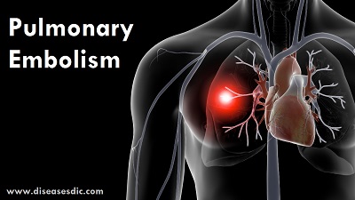
A pulmonary embolism (PE) is a blood clot that develops in a blood vessel in the body (often in the leg). It then travels to a lung artery where it suddenly blocks blood flow.
A blood clot that forms in a blood vessel in one area of the body breaks off, and travels to another area of the body in the blood is called an embolus. An embolus can lodge itself in a blood vessel. This can block the blood supply to a particular organ. This blockage of a blood vessel by an embolus is called an embolism.

Pulmonary Embolism
The heart, arteries, capillaries, and veins make up the body’s circulatory system. Blood is pumped with great force from the heart into the arteries. From there blood flows into the capillaries (tiny blood vessels in the tissues). Blood returns to the heart through the veins. As it moves through the veins back to the heart, blood flow slows. Sometimes this slower blood flow may lead to clot formation.
Epidemiology
The World Health Organization (WHO) estimates a worldwide incidence of PE of 0.75 to 2.69 per 1,000 individuals per year.5 The CDC estimates a rate of 1 to 2 per 1,000 people per year in the United States alone. The mortality rate of acute PE is approximately 7% to 11% and is estimated to cost the United States’ healthcare system $30,000 in the first year after diagnosis.
Risk factors
Factors that increase your risk of developing deep vein thrombosis and pulmonary embolism include:
- Cancer
- A family history of embolisms
- Fractures of the leg or hip
- Hypercoagulable states or genetic blood clotting disorders, including Factor V Leiden, prothrombin gene mutation, and elevated levels of homocysteine
- A history of heart attack or stroke
- Major surgery
- Obesity
- A sedentary lifestyle
- Age over 60 years
- Taking estrogen or testosterone
Causes of Pulmonary embolism
Pulmonary embolism is caused by a blocked artery in the lungs. The most common cause of such a blockage is a blood clot that forms in a deep vein in the leg and travels to the lungs, where it gets lodged in a smaller lung artery.
Almost all blood clots that cause pulmonary embolism are formed in the deep leg veins. Clots also can form in the deep veins of the arms or pelvis.
Sometimes blood clots form in surface veins. But these clots rarely lead to pulmonary embolism.
In rare cases, pulmonary embolism may be caused by other substances. They include:
- Small masses of infectious material.
- It can be released into the bloodstream after some types of bone fractures, surgery, trauma, or severe burns.
- Air bubbles or substances that get into the blood from trauma, surgery, or medical procedures.
- Tumors caused by rapidly growing cancer cells.
- Amniotic fluid.
Symptoms of Pulmonary Embolism
Symptoms usually begin suddenly and may include:
- Sudden shortness of breath
- Sharp chest pain, often aggravated by coughing or movement
- Pain in your back
- Cough with or without bloody sputum.
- Excessive sweating
- Rapid pulse or breathing
- Lightheadedness or passing out
- Blue lips or nail beds
If you have recently had a blood clot in a leg or arm, you may experience:
- Swelling of the affected leg or arm.
- Leg pain or tenderness that may only occur when you are standing or walking.
- Increased warmth in the swollen or painful area of the affected leg or arm.
- Redness or discoloration of your skin.
- Enlargement of superficial veins in the affected leg or arm.
Complications
Pulmonary embolism is a very serious condition that can:
- Cause heart damage.
- Damage part of the lung because of the lack of blood flow to lung tissue, which can lead to pulmonary hypertension.
- Cause low oxygen levels in the blood.
- Damage other organs in the body because of a lack of oxygen.
- Cause death if the blood clot gets too large or if there are multiple blood clots.
Diagnosis and test
Pulmonary embolism can be difficult to diagnose because some people have no obvious symptoms while in others symptoms are non-specific. Diagnosis is also difficult in people with underlying heart or lung disease. Pulmonary embolism is usually diagnosed by one or more of the following tests.
Blood tests
A blood test can reveal a low level of oxygen in the blood, which can be caused by a clot in a lung blood vessel, or high levels of the clot-dissolving substance, D dimer, which may be increased in the presence of blood clots. A blood test can also determine whether someone has an inherited clotting disorder.
Chest x-ray
An x-ray produces film images of the heart and lungs. It cannot diagnose pulmonary embolism but can be used to exclude other causes of chest pain in patients with suspected pulmonary embolism.
Duplex ultrasound
Ultrasound uses sound waves that bounce off blood vessels, which are then converted by a computer into images. Duplex ultrasonography is a type of ultrasound that measures how blood flows through arteries and veins. It is used to check for the presence of blood clots in leg veins.
Spiral computed tomography (CT) scan
This type of CT scan involves a scanner rotating around the body in a spiral to create 3-D images. Spiral CT can detect abnormalities within lung arteries with high precision. Contrast material may be administered by injection into a vein (ie: intravenously) during the CT scan to enhance the outline of the pulmonary arteries.
Ventilation-perfusion (VQ) Scan
During a VQ scan the patient inhales a special aerosol and a special dye is injected into a vein. The aerosol and dye can be seen on x-ray as they move through the lungs. A series of x-rays are then taken, which assess the flow of blood and air through the lungs. Abnormalities may suggest a pulmonary embolism.
Pulmonary angiogram
A pulmonary angiogram is the most accurate way to diagnose pulmonary embolism. A catheter is inserted into a large vein (usually one in the groin) and threaded through into the heart and then into the pulmonary arteries. A special dye is injected and x-rays are taken as the dye travels through the lung arteries, thus providing a clear picture of the blood flow.
A pulmonary angiogram is usually performed when other tests fail to provide a definitive diagnosis. It requires a high level of skill and has potentially serious risks, eg: the dye may cause kidney damage in people with reduced kidney function.
Magnetic resonance imaging (MRI)
MRI involves the use of radio waves and a magnetic field to produce detailed images of the inside of the body. MRI is usually used for pregnant women (to avoid the baby being exposed to radiation) or people whose kidneys may be harmed by dyes or contrast materials that are used in other tests.
Treatment and medications
Treatment is aimed at keeping the blood clot from getting bigger and preventing new clots from forming. Prompt treatment is essential to prevent serious complications or death.
Medications
Blood thinners (anticoagulants): These drugs prevent new clots from forming while your body works to break up the clots. Heparin is a frequently used anticoagulant that can be given through the vein or injected under the skin. It acts quickly and is often overlapped for several days with an oral anticoagulant, such as warfarin, until it becomes effective, which can take days.
A newer class of anticoagulants, referred to as novel oral anticoagulants (NOACs), has been tested and approved for the treatment of venous thromboembolism, including pulmonary embolism. These medications work quickly and have fewer interactions with other medications. Some NOACs have the advantage of being given by mouth, without the need for overlap with heparin. However, all anticoagulants have side effects, with bleeding being the most common.
Clot dissolvers (thrombolytics): While clots usually dissolve on their own, there are medications given through the vein that can dissolve clots quickly. Because these clot-busting drugs can cause sudden and severe bleeding, they usually are reserved for life-threatening situations.
Surgical and other procedures
Clot removal: If you have a very large, life-threatening clot in your lung, your doctor may suggest removing it via a thin, flexible tube (catheter) threaded through your blood vessels.
Vein filter: A catheter can also be used to position a filter in the body’s main vein called the inferior vena cava that leads from your legs to the right side of your heart. This filter can help keep clots from being carried into your lungs. This procedure is typically reserved for people who can’t take anticoagulant drugs or when anticoagulant drugs don’t work well enough or fast enough. Some filters can be removed when they are no longer needed.
Pulmonary Embolism prevention
Preventing new blood clots can prevent PE. Prevention may include
- Continuing to take blood thinners. It’s also important to get regular checkups with your provider, to make sure that the dosage of your medicines is working to prevent blood clots but not causing bleeding.
- Heart-healthy lifestyle changes, such as heart-healthy eating, exercise, and, if you smoke, quitting smoking
- Using compression stockings to prevent deep vein thrombosis (DVT)
- Moving your legs when sitting for long periods of time (such as on long trips)
- Moving around as soon as possible after surgery or being confined to a bed