Description
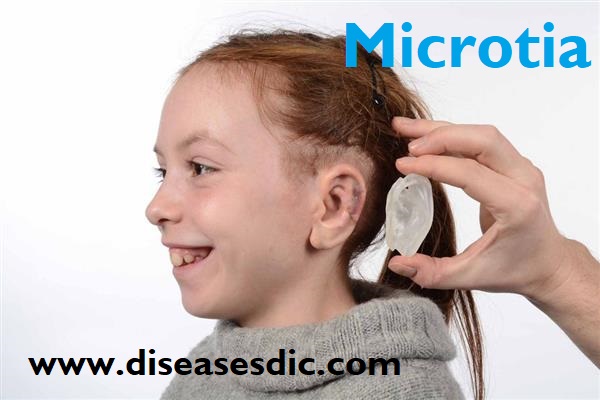
Microtia is a congenital deformity of the outer ear where the ear does not fully develop during the first trimester of pregnancy. The word “microtia” comes from the Latin words “micro” and “otia”, meaning “little ear.” Microtia ears can vary in appearance but are usually smaller in size, often only consisting of a tiny peanut-shaped lobe.
Microtia usually happens during the first few weeks of pregnancy. These defects can vary from being barely noticeable to being a major problem with how the ear formed. Most of the time, microtia affects how the baby’s ear looks, but usually, the parts of the ear inside the head (the inner ear) are not affected. However, some babies with this defect also will have a narrow or missing ear canal.
Grades of microtia
- Grade 1: The ear is smaller than normal but the key features of the normal ear are present, though they may have minor alterations in shape or form.
- Grade 2: Some of the features of the ear are missing, though usually much of the lower two-thirds of the ear is still present. Grade 2 microtia is sometimes called “conchal type microtia.” The ear canal may be present but frequently is very narrow (canal stenosis).
- Grade 3: This is the most common type of microtia, in which the only feature remaining is a small peanut-shaped remnant earlobe. Grade 3 microtia is sometimes called “lobular type microtia.” The ear canal is usually completely absent (aural atresia).
- Grade 4: Complete absence of the external ear without any remnant. This is called “anotia” and is rarely seen.
Pathophysiology
A review of embryology allows a better understanding of the pathophysiology of microtia. The anatomy of the microtic ear is similar to the anlage seen in the 6-week embryo. Microtia often is associated with atresia or absence of the external auditory meatus, suggesting an arrest of development.
The external and middle ear develops from the first (mandibular) and second (hyoid) branchial arches. The auricle begins development at 5 weeks from 6 hillocks, 3 on either side of the first branchial cleft between these 2 arches, which becomes the external canal. Ultimately, the hillocks of the first arch contribute to the tragus and the root of the helix, and the remainder of the auricle develops from the hillocks of the second arch. The middle ear ossicles develop from the first and second arch with the mastoid air cells, eustachian tube, and remaining middle ear developing from the first pharyngeal pouch. The tympanic membrane develops where the first pouch meets the first cleft.
Initially, the ear has a ventromedial position, which becomes more dorsolateral as the midface and mandibular processes grow and push it outward and upward. An interruption in the proliferation or fusion of the hillocks at varying stages of development can produce the variable rudimentary structures that present as microtia.
Causes of Microtia
- Currently, no specific gene has been identified to cause this condition. Several medications have been associated with microtia, but this is hard to prove. One hypothesis is that a small blood vessel (stapedial artery) obliterates or bleeds near the developing ear, causing a decreased flow of cells important in ear development.
- Microtia can occur by itself, or it may occur with syndromes such as hemifacial microsomia, where the entire half of the face is smaller, Treacher Collins syndrome and Goldenhar syndrome.
- It occurs more commonly in infants of diabetic mothers, and infants who were exposed to isotretinoin, thalidomide, alcohol or mycophenolate.
Risk factors
- Diabetes – Women who have diabetes before they get pregnant have been shown to be more at risk of having a baby with anotia/microtia, compared to women who did not have diabetes.
- Maternal diet – Pregnant women who eat a diet lower in carbohydrates and folic acid might have an increased risk of having a baby with microtia, compared to all other pregnant women.
Symptoms of Microtia
If your child has microtia, they might also experience:
- Reduced hearing (by roughly 40 percent) in the affected ear.
- Trouble figuring out which direction a sound is coming from.
- Frequent ear infections.
- The external ear that is absent, or closed (also known as microtia atresia).
Complications of Microtia
- Hearing loss. Beyond the apparent visual deformity of the ear, children with microtia often experience some hearing loss due to the closure or absence of the external ear canal. This hearing loss can affect how the child’s speech will develop.
- Ear infections. Children with microtia may also have ear infections.
- Self-consciousness. Even if a child doesn’t suffer hearing loss, feelings of self-consciousness may develop. Children usually become aware of differences in their bodies around ages 4 or 5. By the age of 6 or 7, children may tease other children who do not look “normal.” The child with microtia may feel self-conscious and inadequate.
Diagnosis and test
Imaging Studies
Microtia is rarely noticed on prenatal ultrasonography, primarily because of the complexity of the fetal ear and the inherent nature of conventional two-dimensional (2D) ultrasonography. Some authors suggest the use of three-dimensional (3D) ultrasonography to provide a better examination of the fetal ear for purposes of prenatal diagnosis and genetic counseling.
If a child is born with microtia, there should be an early evaluation of the ear’s organ systems and the child’s hearing. The first consultation with physicians should be shortly after birth, so parents can understand the treatment options for their child.
Unilateral microtia. In unilateral cases, children generally retain full hearing in the unaffected ear, while still retaining some hearing on the affected side. Even if the ear canal is closed, sound can be absorbed into the still-functioning inner ear. An audiogram (link to glossary) should be performed within the first few months of life. This test should be repeated at yearly intervals. A radiologist will evaluate the middle ear structures and ear canal using a CT scan (link to glossary).
Bilateral microtia. In bilateral cases, children are at risk of greater hearing loss, which can lead to poor speech development. Children with bilateral microtia require earlier intervention. An audiogram is recommended within the first few days of life, ideally before the patient is discharged from the hospital after birth; the test should be repeated until consistent results are obtained. A CT scan will evaluate whether the middle ear structures can be surgically reconstructed. Children with bilateral microtia often use bone-conducting hearing aids before undergoing surgical treatment.
Treatment and therapy
All the treatment options available are cosmetic. That is, they improve the look of the ear but cannot improve its function. There are three options for treatment available:
- No surgical treatment
- Ear reconstruction using a rib graft
- False (prosthetic) ear
More information about each option follows. The team will discuss each option with the family, and the final decision about which option to take will be made jointly between them, their child and the microtia team.
No surgical treatment
While the child is young, parents may decide to leave the ear as it is. Some children camouflage their ear by growing their hair long, while others deal with comments and questions more easily. The chances of successful surgery or prosthesis improve with age, so if the child changes his or her mind in the future, these options will still be open.
Ear reconstruction using a rib graft
This involves making a new ear from a child’s own tissue. The framework or ‘scaffolding’ is made from cartilage taken from their ribcage. The reconstruction is carried out in two stages. It is important that the cartilage is mature so this option is not offered to children under ten years old. Most children are at secondary school when this option is offered.
Rib graft used for the surgery
False (prosthetic) ear
This involves attaching implants and fixtures to the child’s head onto which a false ear is attached. This can be carried out on children around seven years old or more. It usually takes two operations: one to insert the implants and another to attach the fixtures. Once the operations are complete, the false ear is attached to the fixtures. The false ear itself is made from soft, silicone material and is sculpted to match the child’s other ear. It will need to be replaced every two years and the fixtures need to be cleaned every day.
Prevention of Microtia
There is no way to prevent this deformity as it is due to genetic mutation. Thorough genetic test during the pregnancy is advised.

