Definition
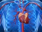
The heart valve disease is the damage in one of the heart valves such as the mitral valve, aortic, tricuspid and pulmonary. It can disrupt the blood flow to the heart by tissue flopping. The heart valves work by ensuring that blood flows in a forward direction and doesn’t back up or cause leakage.
If you have a heart valve disorder, the valve isn’t able to do this job properly. This can be caused by a leakage of blood, which is called regurgitation, a narrowing of the valve opening, which is called stenosis, or a combination of regurgitation and stenosis. Your heart valve disease treatment depends on the heart valve affected and the type and severity of the valve disease. Sometimes heart valve disease requires surgery to repair or replace the heart valve.
Epidemiology
Valvular heart disease (VHD) is a common condition in clinical practices that are strongly connected to heart dysfunction and death. This explains the important changes in the presentation of valvular disease, which now mainly affects predominantly older people. However, rheumatic heart disease remains the main etiology in developing countries.
The overall VHD prevalence in the USA is 2.5% with a wide age-related variation from 0.7–13.3%6. The prevalence increased significantly with age, from less than 2% before 65 years, to 8.5% between 65 years and 75 years, and 13.2% after 75 years. Similar age tendencies were also demonstrated in the Euro Heart Survey7.
Types
There are two main types of heart valve disease
Valvular insufficiency: It is also called regurgitation, incompetence or “leaky valve”. It occurs when the valves are not tight. So the blood may leak across the valve and it may worsen by working harder the valve and the rest blood may flow to the rest of the body. Based on the wall affected it is called as tricuspid regurgitation, pulmonary regurgitation, mitral regurgitation or aortic regurgitation.
Valvular stenosis: It occurs when the valve is smaller than normal due to fused leaflets or stiff. The narrowed valve makes the heart to pump very hard to make blood flow to the body. This leads to heart failure. All the valves can be stenotic and it is called tricuspid stenosis, pulmonic stenosis, mitral stenosis or aortic stenosis.
Risk factors
Some of the risk factors are as follows:
- As older the age your heart valve becomes thicker and stiffer
- Infection of pericardium and lining of heart valves. It is called as Infective Endocarditis
- Congenital heart disorders.
- Myocardial infarction, coronary artery disease, and heart attack
- People who with rheumatic fever, heart failure and previous heart valve diseases are higher risks of heart valve disease
- High blood pressure, smoking, high blood cholesterol, overweight or obesity, insulin resistance and intravenous drug use
- Some babies born with aortic valve had two valves instead of three valves
Causes of heart valve disease
- Heart valve disease can develop before birth or sometimes after the birth can be acquired due to infection and other heart conditions. The main cause of the heart valve disease is unknown.
- Your heart has four valves that make the blood to flow incorrect directions. These valves include the pulmonary valve, aortic valve, tricuspid valve and mitral valve. Each has flaps that close and open when flows in each heartbeat.
- Sometimes infection may also cause heart valve disease. The microbes may enter the bloodstream and thus it infects the surface of the heart muscles and valves. This rare but serious infection is called infective endocarditis. The germs can enter the bloodstream through needles, syringes and other medical devices or through skin wounds or gums.
- Rheumatic fever, untreated strep throat or other infections that progress to rheumatic fever can also cause heart valve disease.
Heart valve diseases include:
Stenosis is the condition when the heart valves become stiff and fused. This may result in narrowing valve opening and decreased blood flow.
Atresia is the condition in the valve is not formed, in which solid sheet of tissue blocks the blood flow between the chambers.
Regurgitation is the condition in which the valves don’t close properly and thus it causes blood to flow backward in your heart. It commonly occurs due to the valve bulging back and the condition is called prolapse.
Heart failure means your heart is working less efficiently and cannot pump a normal amount of blood.
Atrial fibrillation is an abnormal heart rhythm that starts in the atria (upper chambers of the heart). It can cause a rapid, disorganized heartbeat.
Mitral valve prolapse is a common cause of a heart murmur caused by a “leaky” heart valve.
Symptoms associated with heart valve diseases
- Shortness of breath or difficulty catching your breath
- Heart palpitations may come as irregular heartbeats, rapid heart rhythm, skipped beats or a flip-flop feeling in the chest.
- Edema: swelling of ankle, abdomen, feet, and even belly, which can cause you to feel bloated.
- Weakness or dizziness: Feeling very much weak and sick. Sometimes dizziness and fainting can occur.
- Weight can be increased about one or two pounds a day.
- Feels discomfort like pressure or a weight on the chest while doing activities.
- Panic, anxiety, and fatigue.
- Numbness or tingling in the hands and feet.
- Wet cough.
- Blood clots.
Diagnosis and Test
If any people admitted with valvular disease, a doctor will look for patient’s medical history and their physical examination. Some of the diagnostic techniques are also used as follows.
Electrocardiogram (EKG or ECG): It is a diagnosing technique to read about the heart electrical activity, heart rate, rhythm and the size of the heart chambers. It is a painless and non-invasive procedure using electrodes that are attached to various parts of the chest and the trunk region.
Cardiac catheterization: It is fully an invasive procedure, in which a catheter is inserted into an artery present in the leg or arm and slowly propelling into the heart. After that, along with the catheter, a dye is also injected to visualize the damaged heart valves through an X-ray imaging.
Echocardiogram: Sound waves are used to frame a moving image of the heart. The echocardiogram is much more definite than the images obtained by X-ray imaging and also it doesn’t use any radiation. The image obtained from this imaging technique is probably used to find out the deformity of the muscles, valves of the heart. It is also supposed to identify any fluid that is surrounding the heart.
Chest x-ray: it is a technique used electromagnetic radiations to take a picture of the bones. But it is employed to find the changes in the size of the heart. Enlargement of the heart can be easily identified through an X-ray image.
Stress test: Variety of exercise test is performed to identify the amount of stress tolerance and at the same time to monitor the response of the heart to physical exercises. In some cases, patients aren’t able to perform physical exercise, during such conditions medications are used to mimic the effect of exercise on the heart.
Treatment and medication
Heart valve repair
If any of the heart valves is repaired you will probably have any of the following procedures according to the fault in your heart.
- Commissurotomy: it is a procedure to remove the severely narrowed valves because of scar tissue in the flaps of valves, and calcium deposits on the valve. It is usually employed in people shouldn’t have balloon valvotomy.
- Decalcification: Calcium deposits are excised to allow the valve leaflets more flexible and to close free flow.
- Leaflet reshaping: floppy leaflets are excised out, after that the flap will be tailored back together. This allows the valves to close tightly. This procedure is also called as quadrangular resection.
- Chordal transfer: If the anterior leaflet of your mitral valve is floppy (your doctor may say it has prolapse), the tendons that connect your valves — called the chordae — are moved from your posterior leaflet to your anterior leaflet. Then, the posterior leaflet is fixed by the reshape leaflets procedure.
- Annulus support or annuloplasty: The tissue that supports valves called valve annulus. If this valve annulus is too wide then a doctor will tailor a ring-like structure around the valve. The ring may be made of synthetic material or a biological tissue, which maintains the original shape of the valve.
- Patched leaflets: Your surgeon may use tissue grafts to repair any leaflets that have tears or holes.
Heart valve replacement
If the heart valve repair surgery is failed or cannot be repaired, your doctor will go for valve replacement surgery. During this surgery damaged valve is removed and a new synthetic valve is sewn with the tissues of the old valve.
The new valve can be of two varieties as follows:
- Mechanical valve: It is fully made up of mechanical parts, which operates on the external source or automatically. Usually, these valves are covered by a polyester knit fabric.
- Biological or bioprosthetic valve: it is usually made up of animal or human tissue and even it can be generated using natural materials like collagen, chitosan, and other biomaterials through tissue engineering techniques.
Medications
- ACE inhibitors & Vasodilator: As the name suggests it opens the blood vessels fully and can help to reduce high blood pressure and slow heart failure.
- Anti-arrhythmic medications: They help to restore a usual pumping rhythm to the heart.
- Antibiotics: It can help to prevent the occurrence of infections in the heart.
- Anticoagulants (blood thinners): Reduces the risk of developing blood clots due to poor circulation of blood through the faulty heart valves. Blood clots are highly dangerous because they can lead to stroke.
- Beta-blockers: They can reduce the stress by aiding the heart to beat slowly. In some instances, it may help to get rid of the heart palpitations.
- Diuretics (water pill): They assist to reduce the amount of fluid in the tissues and bloodstream which can minimize the stress on the heart.
Lifestyle changes to prevent the onset of heart valve diseases
Healthy lifestyle improves the overall health of the heart and can help to slow the progression of heart diseases. Some healthy choices include:
- Healthy Diet: Take low fat, low salt, low cholesterol diet and also avoid excessive intake of caffeine and alcohol.
- Don’t Smoke: If you do smoke, get help from a doctor to quit. You will immediately escape from the risk of heart disease as soon as you quit
- Reduce mental stress through exercise: Daily physical activity is a great way to get rid of stress, disturbed sleep, excess weight, and also improves your overall sense of wellbeing. Always consult with a doctor before beginning any new physical activity.

