


Understanding Takayasu’s arteritis
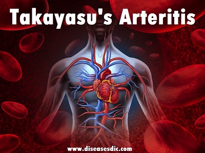
Takayasu’s arteritis is an uncommon form of vasculitis. Inflammation damages large and medium-sized blood vessels. The vessels most commonly affected are the branches of the aorta (the main blood vessel that leaves the heart), including the blood vessels that supply blood to the arms and travel through the neck to provide blood to the brain. The aorta itself is also often affected.

Segments of blood vessels can weaken and stretch, resulting in an aneurysm. They can also become inflamed and narrowed, resulting in restricted blood flow. Lastly, blood vessels can become completely blocked (called an occlusion).

Less commonly, arteries that provide blood flow to the heart, intestines, kidneys and legs may be involved. Inflammation of large blood vessels may cause segments of the vessels to weaken and stretch, resulting in an aneurysm. Vessels can also become narrowed or even completely blocked (called an occlusion).
Takayasu’s arteritis Prevalence
Takayasu’s arteritis is primarily a disease of adolescent girls and young women, who account for 80-90% of cases. The prevalence is highest in the Asian population but it is seen in all races and geographic areas. South America has now been recognized as an area of high incidence.
Types
Takayasu’s arteritis can be divided into the following six types based on angiographic involvement.
Type I – Branches of the aortic arch
Type IIa – Ascending aorta, aortic arch, and its branches
Type IIb – Type IIa region plus thoracic descending aorta
Type III – Thoracic descending aorta, abdominal aorta, renal arteries, or a combination
Type IV – Abdominal aorta, renal arteries, or both
Type V – Entire aorta and its branches
Takayasu’s arteritis Risk factors
Common risk factors in the development of Takayasu’s arteritis include:
- Female gender
- Age <40
- Asian ethnicity
- Genetic predisposition
Infectious agents such as:
- Spirochetes
- Mycobacterium tuberculosis
- Streptococcal organisms
Causes of Takayasu’s arteritis
In Takayasu’s arteritis, the aorta and other major arteries, including those leading to the head and kidneys, become inflamed. Over time, the inflammation causes changes in these arteries, including thickening, narrowing and scarring.
The result is reduced blood flow to vital tissues and organs, which can lead to serious complications and even death. Sometimes arteries become abnormally dilated, leading to aneurysms that may rupture.
Just what causes the initial inflammation in Takayasu’s arteritis isn’t known. It’s likely that Takayasu’s arteritis is an autoimmune disease in which the immune system malfunctions and attacks own arteries as if they were foreign substances. The disease may be triggered by a virus or other infection.
Takayasu’s arteritis Symptoms
Symptoms vary depending on which arteries have become narrow. As with many other inflammatory diseases, patients with Takayasu’s arteritis often feel ill with “flu-like” symptoms before the disease is treated. Symptoms include:
- Pain in the hand or lower leg with use (claudication), often with absent pulse or abnormally reduced blood pressure
- Abdominal pain especially after eating
- Chest pain with activity
- Dizziness
- Headache due to very high blood pressure (caused by narrowing of an artery supplying a kidney)
- Muscle and joint pain, fatigue, fever

Takayasu’s arteritis Complications
Even with treatment, damage to the aorta may be permanent. Repeated swelling and healing of arteries can lead to complications. Therefore, it is important to follow up with your doctor on a regular basis. Complications can include:
- Hardening and narrowing of the blood vessels
- High blood pressure
- Heart valve disorder (eg, aortic valve damage)
- Heart failure
- Aortic aneurysm
- Transient ischemic attack (TIA), a “mini-stroke” that doesn’t cause permanent damage but serves as a warning sign
- Stroke
- Heart attack
- Pulmonary artery problems
Pregnancy: Successful pregnancies are possible for patients with TAK. If you are pregnant, or planning to become pregnant, talk to your doctor about potential effects of the disease and treatments.
Diagnosis and test
Blood Tests: There is no blood test that can be used to definitively confirm the diagnosis. The erythrocyte sedimentation rate (ESR) and C-reactive protein (CRP) can help measure activity of the illness and your response to treatment.
Imaging: Imaging studies are useful in the diagnosis and monitoring of TA. Their use may allow diagnosis before arterial damage is severe. However, none of them are 100 per cent accurate.

An abnormal aortic arch in a patient with Takayasu’s arteritis
Magnetic resonance imaging (MRI) is commonly used to aid diagnosis and assess the extent of TA. The scan does not involve radiation and the picture of the arteries can be enhanced by using a contrast medium (that only rarely causes an allergic reaction).
MRI is a safe means by which to monitor the progress of disease and response to treatment. The main drawbacks are that the scanner is very noisy and rather claustrophobic.
Ultrasound – Doppler ultrasound (ultrasound scan): uses a probe which is passed over the carotid arteries in the neck. This allows assessment of blood flow and can detect and monitor the arterial wall and the degree of narrowing.
Positron emission tomography (PET scanning): requires the patient to be injected with radioactive fluorodeoxyglucose. PET scans are particularly good at detecting inflammation in the arterial wall and are often used in combination with CT scanning (CT-PET). PET may identify early active disease and may on occasion help to distinguish active disease (which needs further treatment) from scarring resulting from previous inflammation.
Computed tomography (CT) angiography: Contrast-enhanced CT angiography is useful for the diagnosis of TA and can help identify the extent of damage to the arteries. Unlike MRI/MRA and Doppler US, CT angiography does involve radiation.
Treatment and medications
A main goal of treatment is to reduce damage to your arteries. This is done by taking medicine that reduces inflammation. You may be given medicine such as:
Glucocorticoid medicine: Prednisone is a glucocorticoid medicine that can help treat Takayasu arteritis. Taking this for a long time can damage your bones. Taking calcium and vitamin D supplements may help prevent this damage. This medicine doesn’t work for all people.
Immunosuppressant medicine: This may be given if prednisone doesn’t work for you, or stops working. This medicine suppresses the immune system and helps prevent the inflammation. The medicines include methotrexate and azathioprine. These medicines don’t work for all people.
Anti-tumor necrosis factor (anti-TNF): Etanercept and infliximab are 2 examples of anti-TNF medicine.

Surgery
Your healthcare provider will need to watch the health of your large arteries on a regular basis. You will have blood tests and imaging tests such as MRA or CTA.
If 1 of your arteries becomes very narrow, you may need surgery (called revascularization) to repair it. This surgery can be done with angioplasty or a bypass graft:
- Angioplasty: The artery is made wider.
- Bypass graft: The blood flow is sent around the blocked part of the artery by attaching part of another blood vessel around it.
Prevention of Takayasu’s arteritis
As the precise etiology of Takayasu arteritis and causes of flare-ups in disease activity are unknown, there are no known specific preventive actions. Management of hypertension is important to prevent further vascular damage.
- Attention to osteoporosis screening and management is crucial, given the need for glucocorticoid therapy.
- Patients require influenza and pneumococcal immunizations annually.
- Use of prophylactic antibiotic therapy to prevent Pneumocystis jirovecii pneumonia is important, especially when the prednisone dose is more than 20 mg daily.
- Atherosclerotic vascular disease can further complicate the vascular damage caused by Takayasu arteritis; thus, control of other risk factors is important.