Definition
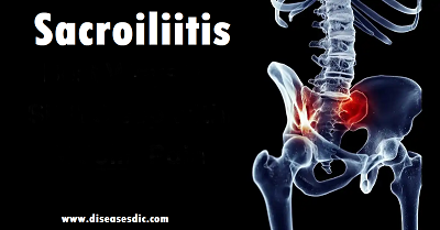
Sacroiliitis is a condition in which the sacroiliac (SI) joint – the large joint located between the sacrum (the tailbone) and the ilium (the pelvis) becomes inflamed or disrupted, and causes pain. There are two sacroiliac joints, one on the right side and one on the left, and they are responsible for transferring weight and forces between your upper body and legs.
Sacroiliitis can be seen after trauma to the pelvis, in individuals with inflammatory disorders such as ankylosing spondylitis, and in patients with a history of previous lumbar surgery. An arthritic sacroiliac joint that is undergoing degeneration is also among the causes of sacroiliitis. In women, postpartum sacroiliac pain after delivery is not uncommon and often resolves spontaneously. However chronic postpartum sacroiliitis may persist and can be quite disabling.
Anatomy of the Sacroiliac Joint
The sacroiliac joint is a highly complex joint and is most often classified as a “diarthrodial” joint. It has several components to its structure:
- Fibrous joint capsule with thick synovial fluid
- Cartilaginous surfaces
- Ligamentous connections.
Unlike most joints, the SIJ is not very mobile – it serves more to provide stability and weight-bearing capability. While there is some small amount of movement in the SIJ, there has not been a relationship seen between the degree of movement of the joint and pain.
The joint is supported by a group of muscles that attach to the joint itself and aid with walking, sitting, standing, and as well as providing support and stability. These muscles include the gluteus maximus and medius, biceps femoris, piriformis, latissimus dorsi and erector spinae. The SIJ is designed for stability and weight-bearing.
The nerve supply to the SIJ is equally as complex and has been the subject of some debate amongst physicians.
- Dorsal Aspect: innervated by the S1-S3 dorsal rami. Some studies suggest innervation from the L5 nerve, and a recent cadaver study found contribution from the S4 nerve in 59% of joints.
- Ventral Aspect: innervated by the L4-S2 ventral rami, while others include levels as high as L2.
Most physicians will agree that pain from the SIJ can occur from within the joint as well as immediately outside it due to pain receptors being located throughout the joint capsule as well as in the adjacent muscles and ligaments. Pain receptors within the joint capsule are mostly found in the proximal and middle thirds of the joint – this is where most procedures targeting pain from the SIJ will be performed.
Types of Sacroiliitis
A variety of conditions and circumstances can cause sacroiliitis. There are many influencers of sacroiliitis, so it is broken down into three types:
- Inflammatory
- Degenerative
- Pyogenic
Inflammatory sacroiliitis happens when your sacroiliac joint tissue becomes inflamed for various reasons that are not degenerative or organic.
Pyogenic sacroiliitis is the joint’s inflammation due to an infection.
Degenerative sacroiliitis forms because a degenerative bone or joint condition caused it.
Epidemiology
Reports on the prevalence of sacroiliac pain vary widely. Some studies report the prevalence as 10% to 25% of those with lower back pain. In those with a confirmed diagnosis, the presentation of pain was ipsilateral buttock (94% cases) and midline lower lumbar area (74%). Up to 50% of cases have radiation to the lower extremity: 6% to the upper lumbar area, 4% percent to the groin, and 2% percent to the lower abdomen. Symmetrical sacroiliitis is found in more than 90% of ankylosing spondylitis and 2/3 in reactive arthritis and psoriatic arthritis.
It is less severe and more likely to be unilateral and asymmetrical in reactive arthritis, psoriatic arthritis, arthritis of chronic inflammatory bowel disease and undifferentiated spondyloarthropathy. The hospital prevalence of sacroiliac diseases is 0,55%, the female sex predominates (82,35%) and the mean age of 25,58 years. Gyneco-obstetric events are the predominant risk factors (47,05%). The etiologie found are bacterial arthritis (82,3%) mainly pyogenic (70,58%), osteoarthritis (11,7%) and ankylosing spondylitis (5,9%).
Pathophysiology
The sacrum articulates with the ilium, which helps to distribute body weight to the pelvis. The SI joint capsule is relatively thin and often develops defects that enable fluid, such as joint effusion or pus, to leak out onto the surrounding structures. As surrounding muscles and structures are affected different presentations of pain can present because different nerve roots innervate these structures. Common distributions of pain are L4-L5 dermatomes, but distributions can certainly present in dermatomes as high as L2 and as low as S3. The asymmetric motion of the pelvis can cause mechanical dysfunction, leading to degeneration and significant pain. Differential diagnosis includes leg-length discrepancies, unilateral weaker limb or gluteal muscles, tight or strained surrounding muscles structures, or hip osteoarthritis.
Causes of Sacroiliitis
Causes for sacroiliac joint dysfunction include:
Traumatic injury: A sudden impact, such as a motor vehicle accident or a fall, can damage your sacroiliac joints.
Arthritis: Wear-and-tear arthritis (osteoarthritis) can occur in sacroiliac joints, as can ankylosing spondylitis – a type of inflammatory arthritis that affects the spine.
Pregnancy: The sacroiliac joints must loosen and stretch to accommodate childbirth. The added weight and altered gait during pregnancy can cause additional stress on these joints and can lead to abnormal wear.
Infection: In rare cases, the sacroiliac joint can become infected.
Sacroiliitis symptoms
Symptoms of sacroiliitis or SI joint pain
- Pain in the lower back
- Stiffness in lower back
- Mild to moderate ache in low back
- Pain in the buttock’s region
- Pain with walking or pain while sitting
- Unilateral low back pain
- Referred pain in lower limb, often mistaken as ‘sciatica’
Symptoms of Sacroiliitis
Risk factors
A history of bone, joint or skin infections: Some people are more prone to infections, and an infection is one possible cause of sacroiliitis.
Injury or trauma to your spine, pelvis or buttocks: Torn ligaments or trauma may create inflammation or infection of the sacroiliac joints.
Urinary tract infection: This infection may spread from your urinary tract, which includes your kidneys, bladder and urethra, to your sacroiliac joints.
Pregnancy: The pelvic bone’s expansion to prepare for childbirth may inflame the area around your sacroiliac joints.
Endocarditis: This infection of your heart’s inner lining may spread to your sacroiliac joints.
Complications
- Left untreated, sacroiliitis causes a loss of mobility for some people.
- Untreated pain also can disrupt your sleep and lead to psychological conditions like depression.
- Sacroiliitis associated with ankylosing spondylitis can progress over time.
- Over time, this type of arthritis causes the vertebrae (bones) in your spine to fuse together and stiffen.
Diagnosis
A high index of suspicion is required to diagnose sacroiliitis as symptoms mimic that of lumbar spine pathologies. A local tenderness directly over sacroiliac joint will elicit pain. Certain specific physical tests to stress Si joint will cause pain on the affected side.
X-ray/MRI/CT Scan: X-ray will show joint arthritis and hardening of bone around SI joint. In infection, joint will be destroyed. MRI will show inflammation and infection in detail. CT scan will show roughened joint surfaces.
Blood Tests: ESR and CRP will be elevated in inflammatory and infectious arthritis. Positive HLA B-27 will help in diagnosing Ankylosing Spondylitis.
Anesthetic Injection: A local anesthetic injection given inside the joint should relieve pain coming from SI joint. It is either done under fluoroscopy guidance in operation theater or under CT guidance.
Treatment for Sacroiliitis
Treatment depends on your signs and symptoms, as well as the cause of your sacroiliitis.
Medications
Depending on the cause of your pain, your doctor might recommend:
- Pain relievers. If over-the-counter pain medications don’t provide enough relief, your doctor may prescribe stronger versions of these drugs.
- Muscle relaxants. Medications such as cyclobenzaprine (Amrix, Fexmid) might help reduce the muscle spasms often associated with sacroiliitis.
- TNF inhibitors. Tumor necrosis factor (TNF) inhibitors – such as etanercept (Enbrel), adalimumab (Humira) and infliximab (Remicade) – often help relieve sacroiliitis that’s associated with ankylosing spondylitis.
Therapy
Your doctor or physical therapist can help you learn range-of-motion and stretching exercises to maintain joint flexibility, and strengthening exercises to make your muscles more stable.
Surgical and other procedures
If other methods haven’t relieved your pain, you doctor might suggest:
- Joint injections. Corticosteroids can be injected into the joint to reduce inflammation and pain. You can get only a few joint injections a year because the steroids can weaken your joint’s bones and tendons.
- Radiofrequency denervation. Radiofrequency energy can damage or destroy the nerve tissue causing your pain.
- Electrical stimulation. Implanting an electrical stimulator into the sacrum might help reduce pain caused by sacroiliitis.
- Joint fusion. Although surgery is rarely used to treat sacroiliitis, fusing the two bones together with metal hardware can sometimes relieve sacroiliitis pain.
Home remedies and exercise
Rest: Avoiding the movements that aggravate sacroiliitis pain can help to reduce inflammation.
Ice and heat: Alternating placing ice and heat packs on the affected area may help relieve sacroiliitis pain. A person should always cover ice and heat packs with a towel to prevent burns and damage to the skin.
Hip flexion exercises: This exercise involves laying on the back with the legs supported by a box or pillows. Cross one leg over the other, squeeze the legs together and then release. Repeat this on both sides. Try a variation of this exercise by laying on the back, lifting the legs, and then squeezing them together with a pillow in between.
Prevention of Sacroiliitis
A positive attitude, regular activity, and a prompt return to work are all very important elements of recovery. If regular job duties cannot be performed initially, modified (light or restricted) duty may be prescribed for a limited time.
Prevention is key to avoiding recurrence:
- Proper lifting techniques
- Good posture during sitting, standing, moving, and sleeping
- Regular exercise with stretching /strengthening
- An ergonomic work area
- Good nutrition, healthy weight, lean body mass
- Stress management and relaxation techniques
- No smoking

