


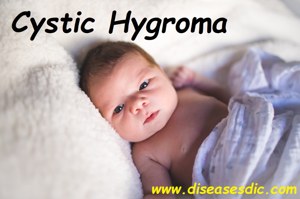
Cystic Hygroma – Description
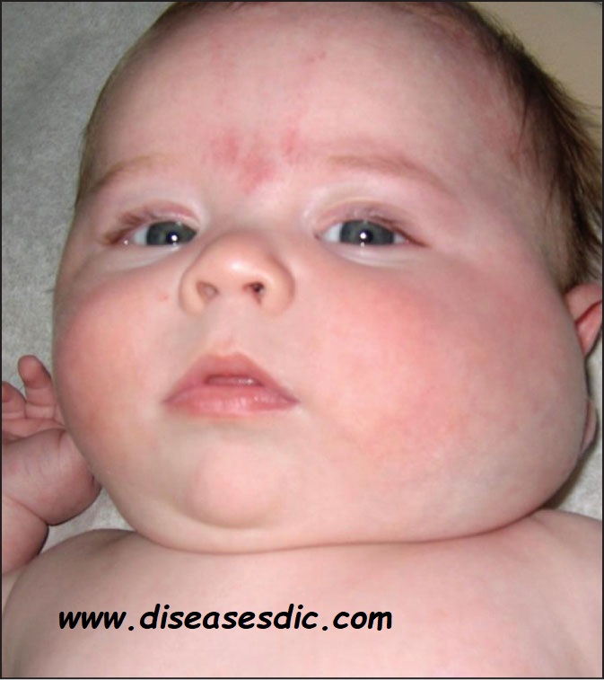
A cystic hygroma is a fluid-filled sac that results from a blockage in the lymphatic system. They are also known as Lymphatic Malformation (LM). It is most commonly located in the neck or head area but can be located anywhere in the body. It may be discovered in a fetus during a pregnancy ultrasound, or it may be apparent at birth as a soft bulge under the skin.

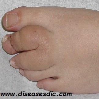
Cystic Hygroma in Foot Fingers

By the end of the fifth week of pregnancy, the baby’s lymphatic tissues form as lymph sacs. The first to appear to serve the chest, arms, neck, and head. They sprout a network of channels called lymphatic vessels that maintain fluid in the baby’s body and carry fats and immune system cells. When a problem occurs between the veins and developing lymph sacs, the sacs expand with fluid and partially or completely block this vessel system.
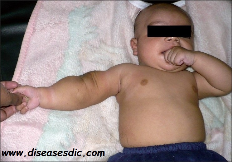
Cystic Hygroma in Elbow

Pathophysiology of Cystic Hygroma
LMs are thought to arise from a combination of the following:
- Failure of lymphatics to connect to the venous system
- Abnormal budding of lymphatic tissue
- Sequestered lymphatic rests that retain their embryonic growth potential
These lymphatic rests can penetrate adjacent structures or dissect along fascial planes and eventually become canalized. These spaces retain their secretions and develop cystic components because of the lack of a venous outflow tract. The nature of the surrounding tissue determines whether the lymphangioma is capillary, cavernous, or cystic.
LMs tend to form in loose areolar tissue, whereas capillary and cavernous forms of lymphangiomas tend to form in muscle. Studies using cell proliferation markers have demonstrated that lymphangioma enlargement is related more to engorgement than to actual cell proliferation. Molecular studies suggest that vascular endothelial growth factor C (VEGF-C) and its receptors may play an important role in the development of LMs.
In addition to congenital development, LMs can be acquired. They can arise from trauma (including surgery), inflammation, or obstruction of a lymphatic drainage pathway.
What Causes Cystic Hygroma?
Cystic hygromas can develop due to genetic disorders or environmental factors. One or more growths may be present at the time of diagnosis.
Common environmental causes of cystic hygromas are:
- Viral infections passed from the mother to the baby during pregnancy
- Exposure to drugs or alcohol during pregnancy
Cystic hygromas are seen more often in infants with genetic diseases. They are particularly common in infants with chromosomal abnormalities. Some genetic conditions associated with hygromas include:
- Turner’s syndrome, in which female children have one X chromosome instead of two
- Trisomy 13, 18, or 21, conditions where children have an extra copy of a chromosome
- Noonan syndrome, a disorder caused by a change (mutation) in one of seven particular genes
Risk factors
The risk factors of Lymphangioma include:
- Genetic disorders, like Turner syndrome, Down syndrome, and Noonan syndrome, are associated with Lymphangiomas
- Any of the Trisomies, such as Trisomy 13 (Patau’s syndrome), Trisomy 18 (Edwards syndrome), and Trisomy 21 (Down syndrome), can increase the risk
- Alcohol abuse
- A viral infection during pregnancy
Signs and Symptoms
Symptoms of a cystic hygroma vary depending on the location of the cysts. Some children may not experience any symptoms other than growth. If a child has symptoms, they may include:
- Fluid-filled sacs on the tongue
- Large cysts that appear blue
- Obstructive sleep apnea, a sleep disorder that causes breathing to stop and start
- Breathing and feeding difficulties
- Failure to thrive
- Bone and teeth abnormalities
In rare cases, the hygromas may bleed or become infected.
Complications During Cystic Hygroma
The possible complications due to Lymphangioma include:
- Infection during the surgical intervention, which may necessitate multiple surgeries
- The growth of the tumor after surgery, due to its incomplete removal
- Adjoining structures, tissues, nerves, and organs, can be damaged during the surgery
- Lymphangioma formations during early stages of pregnancy can complicate the pregnancy and affect normal fetal growth
- Blockage of the food pipe (tracheal or bronchial obstruction), windpipe (esophageal obstruction), or intestine (intestinal obstruction), by the overgrown mass
Diagnosis and test
Prenatal diagnosis
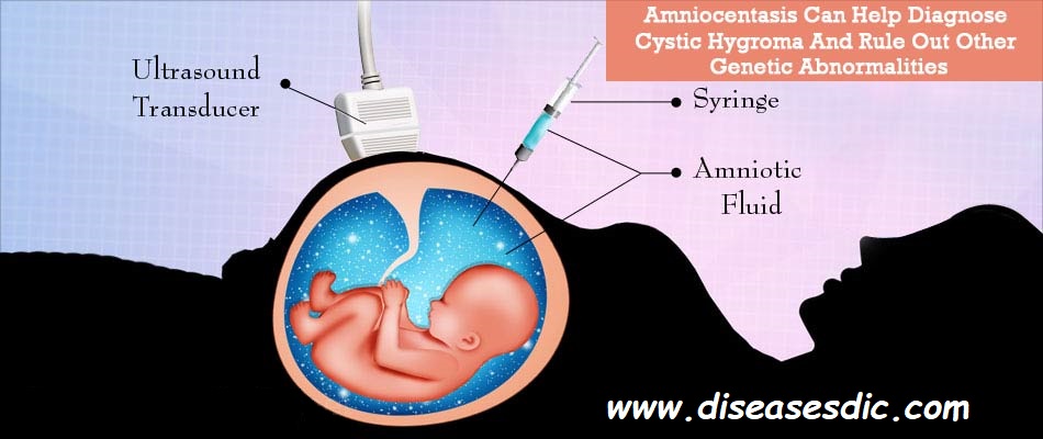
- Occasionally a cystic hygroma in the fetus may be picked up during maternal ultrasound during pregnancy. The doctor may then recommend an amniocentesis test to rule out fetal genetic abnormalities.
- During amniocentesis, a sample of amniotic fluid is obtained by introducing a needle via the pregnant abdomen into the uterus under ultrasound guidance. The amniotic fluid is then tested for genetic abnormalities

Postnatal Diagnosis
This refers to diagnosis after the baby is born.
- Some children have visible lesions which enable readily making a diagnosis on physical examination
- In children where the swelling is not readily visible or thought to compress other structures, these can be demonstrated with imaging investigations such as MRI scan, CT scan, ultrasound, and occasionally even with x-rays
- Other specialized studies such as airway fluoroscopy and lymphoscintigraphy (to outline the lymph network by radiographic imaging) have been occasionally used.
- Rarely endoscopic biopsy to remove a sample of the swelling has been performed for confirmation of diagnosis
Treatment and Prevention of Cystic Lymphangioma
Treatment involves surgical removal of the abnormal tissue whenever possible. However, cystic hygromas can often invade other parts of the neck, making this impossible.
Other treatments have been attempted with only limited success, including:
- Chemotherapy medications
- Injection of sclerosing medications
- Radiation therapy
- Steroids
Let’s see the treatment required for before and after pregnancy
Prenatal Cystic Hygromas
Cystic hygromas diagnosed when the baby is in the womb are not treated. Instead, the doctor will closely monitor the baby’s health. There is an increased risk of miscarriage and intrauterine fetal death. Sometimes prenatal cystic hygromas disappear before birth. If there is spontaneous disappearance of the cystic hygroma by 20 weeks of pregnancy, the chances of chromosomal abnormalities are less.
It is advisable to schedule the delivery in a specialist center to avoid complications during birth
Postnatal Cystic Hygromas
In babies with no symptoms due to the swelling, the doctors may adopt a ‘watch and wait’ approach
Surgery: In babies with symptomatic swelling, surgical removal of the swelling under general anesthesia is performed. The entire swelling should be excised to prevent future recurrences. Surgery requires a hospital stay for a few days.
Sclerotherapy: In larger lesions, sclerotherapy using chemicals to scar or shrink the growth is another method. Once the growth becomes smaller, it may be surgically excised. If growth recurs, multiple sclerotherapy sessions may be necessary. The procedure is performed by an interventional radiologist under general anesthesia. It may be done as a day procedure.
Radiation therapy: Other treatment modalities such as radiofrequency ablation, laser-induced thermotherapy, chemotherapy and steroid medications have also been employed to shrink the lesion
One should not puncture the cyst or drain the swelling without medical supervision. It can lead to severe bleeding or infection.
Complications of Various Treatments
- Bleeding and infection can occur during any invasive procedure
- Surgery – complications related to anesthesia, damage to nerve, muscle or other tissues during surgery
- Sclerotherapy – Allergic reactions to injected chemical, recurrence, scar formation
Outlook
- The long-term outlook for cystic hygromas depends on the size and location of the growth.
- Some cases of cystic hygromas have associations with other genetic conditions that may impact a child’s development.
- Cystic hygromas can return even after treatment or multiple treatments, especially if doctors cannot remove all the tissue.