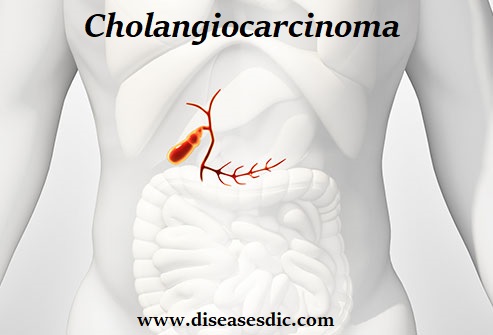
Overview – Cholangiocarcinoma
Cholangiocarcinoma or Bile Duct Cancer is a rare and often fatal cancer that affects the bile ducts. The bile ducts are a series of tubes that transport digestive juices called bile from your liver (where it’s made) to your gallbladder (where it’s stored). From the gallbladder, ducts carry bile to your gut, where it helps to break down fats in the foods you eat. In most cases, cholangiocarcinoma arises in those parts of the bile ducts that lie outside the liver. Rarely, cancer can develop in ducts that are located within the liver.
Cholangiocarcinoma (Bile Duct Cancer) Types
There are several types of cholangiocarcinoma or bile duct cancer. The bile duct is a thin tube that spans from the liver to the first part of the small intestine (duodenum). Its main function is to transport bile, a fluid that aids in the digestion of the fats in foods. Cholangiocarcinoma can develop anywhere within the bile duct system, and the condition is classified based on its site of origin.
Main types of bile duct cancer
The three main types of cholangiocarcinoma are:
- Intrahepatic bile duct cancers – Develop in the small bile duct branches within the liver
- Perihilar bile duct cancers (Klatskin tumors) – Form at the hilum, the point where the hepatic ducts join together to exit the liver
- Distal (extrahepatic) bile duct cancers – Found further down in the bile duct, closer to the duodenum
Cholangiocarcinoma can also be divided by cell types, which are determined based on the appearance of the cancer cells when viewed under a microscope. Most bile duct cancers are adenocarcinomas that start in the glandular cells lining the inside of the duct. Other types of cholangiocarcinoma, such as sarcomas, lymphomas, and small cell cancers, are less common.
Pathophysiology
Cholangiocarcinomas arise from the intrahepatic or extrahepatic biliary epithelium. More than 90% are adenocarcinomas, and the remainder is squamous cell tumors. The etiology of most bile duct cancers remains undetermined. Long-standing inflammation, as with primary sclerosing cholangitis (PSC) or a chronic parasitic infection, has been suggested to play a role by inducing hyperplasia, cellular proliferation, and, ultimately, malignant transformation. Intrahepatic cholangiocarcinoma may be associated with chronic ulcerative colitis and chronic cholecystitis.
Cholangiocarcinomas tend to grow slowly and to infiltrate the walls of the ducts, dissecting along tissue planes. Local extension occurs in the liver, porta hepatis, and regional lymph nodes of the celiac and pancreaticoduodenal chains. Life-threatening infection (cholangitis) may occur that requires immediate antibiotic intervention and aggressive biliary drainage.
Causes of bile duct cancer
The cause of bile duct cancer is unknown. Most happen without a known cause, although some things can increase your risk of getting it.
These include:
- Primary sclerosing cholangitis – a rare type of liver disease that causes long-term inflammation of the liver
- Bile duct abnormalities – such as cysts (fluid-filled sacs) in the bile ducts that are present from birth
- Biliary stones in the liver – hard stones, similar to gallstones, that form in the bile ducts
- Infection from a liver fluke parasite (a parasitic worm; usually only a problem in Asian countries)
- Exposure to certain chemicals and toxins, including thorotrast (a type of dye that used to be used in medical scans)
There may also be a link between long-term hepatitis B and hepatitis C infections, liver scarring (cirrhosis), diabetes, obesity, smoking, and excessive alcohol consumption.
Risk factors
Factors that may increase your risk of cholangiocarcinoma include:
- Primary sclerosing cholangitis. This disease causes hardening and scarring of the bile ducts.
- Chronic liver disease. The scarring of the liver caused by a history of chronic liver disease increases the risk of cholangiocarcinoma.
- Bile duct problems present at birth. People born with a choledochal cyst, which causes dilated and irregular bile ducts, have an increased risk of cholangiocarcinoma.
- A liver parasite. In areas of Southeast Asia, cholangiocarcinoma is associated with liver fluke infection, which can occur from eating raw or undercooked fish.
- Older age. Cholangiocarcinoma occurs most often in adults over age 50.
- Smoking is associated with an increased risk of cholangiocarcinoma.
Symptoms of bile duct cancer
There are not usually any symptoms of bile duct cancer until it grows large enough to block the bile ducts.
This can cause:
- Yellowing of the skin and whites of the eyes (jaundice)
- Itchy skin
- Your poo to turn pale
- Darkening of your pee
- Loss of appetite and weight loss
- Persistent tiredness and feeling unwell
- Tummy (abdominal) pain and swelling – some people feel a dull ache in the upper right side of their abdomen
- High temperature
- Chills and shivering
See your GP if you have persistent symptoms that you’re worried about – particularly if you have jaundice. These symptoms can have several causes, so it’s important to get a proper diagnosis.
What are the complications of bile duct cancer?
Obstruction of the bile duct can lead to infection of the bile drainage system or cholangitis.
Cirrhosis may develop in bile duct cancer. This may be due to the tumor obstructing the bile duct and causing liver cell destruction and scarring. This is especially true in patients with primary sclerosing cholangitis. Both cirrhosis and sclerosing cholangitis are listed as potential risk factors for bile duct cancer.
Other complications may be a consequence of the procedures used to diagnose and treat cancer. These include complications of surgery, chemotherapy, and radiation therapy.
Bile Duct Cancer (Cholangiocarcinoma): Diagnosis
This section describes options for diagnosing bile duct cancer. Not all tests listed below will be used for every person. Your doctor may consider these factors when choosing a diagnostic test:
- The type of cancer suspected
- Your signs and symptoms
- Your age and general health
- The results of earlier medical tests
In addition to a physical examination, the following lab tests may be used to diagnose bile duct cancer or identify it as the cause of jaundice:
Blood chemistry tests. Blood chemistry tests measure the levels of bilirubin and alkaline phosphatase and check other liver functions. High levels of these substances could indicate that the bile duct is not working well.
Tumor marker tests (CEA and CA19-9). Tumor marker tests look for higher-than-normal amounts of certain substances in the blood, urine, or tissues of people with certain types of cancer. Bile duct cancer may cause high levels of carcinoembryonic antigen (CEA) and CA19-9 in the blood. However, a person can have bile duct cancer even if there are normal levels of these tumor markers. Also, there are diseases other than cancer that sometimes cause high levels of these substances.
Other tests may be performed to provide more information about bile duct cancer. These include:
Biopsy. A biopsy is the removal of a small amount of tissue for examination under a microscope. A pathologist then analyzes the sample(s). A pathologist is a doctor who specializes in interpreting laboratory tests and evaluating cells, tissues, and organs to diagnose disease.
The type of biopsy performed depends on the location of the tumor. The doctor can remove tissue samples during a procedure called a percutaneous transhepatic cholangiography (PTC), which is now rarely used, or another procedure called an endoscopic retrograde cholangiopancreatography (ERCP). PTC and ERCP are described below. Or, a computed tomography scan (CT or CAT scan, see below) may help guide a thin needle through the skin into the area to collect a sample of cells.
Ultrasound. An ultrasound uses sound waves to create a picture of the internal organs. During an ultrasound, the doctor may be able to see the actual tumor. However, more often, the ultrasound will show that the small bile ducts have become larger. This is called a “dilation of ducts.” The small bile ducts are located behind a blockage of 1 of the larger bile ducts. Ultrasound can be used to guide a needle through the skin and into the liver toward a suspected cancer to get a sample tissue for diagnosis.
Computed tomography (CT or CAT) scan. A CT scan takes pictures of the inside of the body using x-rays taken from different angles. A computer combines these pictures into a detailed 3-dimensional image that shows any abnormalities or tumors. A CT scan can be used to measure the tumor’s size. Sometimes, a special dye called a contrast medium is given before the scan to provide better detail on the image. This dye can be injected into a patient’s vein or given as a pill or liquid to swallow. A CT scan can also be used to guide a needle to a suspected tumor.
Magnetic resonance imaging (MRI). An MRI uses magnetic fields, not x-rays, to produce detailed images of the body. A specialized MRI used for the bile duct is called MRI cholangiopancreatography. A contrast medium may be given before the MRI to create a clearer picture. This dye can be injected into a patient’s vein or given as a pill or liquid.
PTC. During this test, a thin needle is inserted into the bile duct in the liver. The doctor injects a contrast medium through the needle that allows the bile ducts to show up on x-rays. By looking at the x-rays, the doctor can tell whether and where there is a blockage of the bile ducts. This is important in planning treatment.
Laparoscopy. During laparoscopy, a doctor views the bile duct, gallbladder, and liver through a lighted tube, called a laparoscope. The laparoscope is inserted into a surgical opening in the person’s abdomen. Some laparoscopes can help the doctor take a tissue sample by using small instruments through the tube.
Bile Duct Cancer Treatment
You might have one or more of these treatments:
Biliary drainage. If your bile duct is blocked, your doctor may do a bypass, cutting it off and reattaching it on the other side of the blockage. They could also put a tube called a stent into the duct to keep bile flowing.
Photodynamic therapy. Your doctor injects an inactive form of a certain medication into your vein. The drug tends to collect in more cancer cells than healthy cells. After a few days, they use an endoscope to aim for a special light at the tumor. It activates the drug, killing the cancer cells.
Radiation. This uses high-energy rays or particles to kill cancer cells. Your doctor may use it before surgery to shrink a tumor. After surgery, it can kill any cancer cells that remain. If your doctor can’t remove cancer but it hasn’t spread, radiation can help keep it under control.
Chemotherapy. As with radiation, doctors often use medicines to kill cancer cells before or after surgery. You can take chemo by mouth or through a shot into a vein.
Surgery. Depending on cancer’s spread, your doctor might need to remove some or all of your bile duct, lymph nodes, liver, pancreas, or small intestine.
Liver transplant. This is a rare treat that can sometimes cure bile duct cancer. Your doctor might use chemo and radiation while you wait for a new liver.
Prevention
Different factors cause different types of cancer. Researchers continue to look into what factors cause bile duct cancer, including ways to prevent it. Although there is no proven way to completely prevent this disease, you may be able to lower your risk.
Avoid coming in contact with hazardous chemicals. Thorium dioxide, a chemical once used in x-ray examinations, is associated with a high risk of developing bile duct cancer. Its use has been banned. However, other hazardous chemicals are still available or found in the environment that can increase the risk of developing bile duct cancer.
Avoid alcohol abuse, which can lead to cirrhosis.
If you travel to parts of the world where liver flukes are common, drink only purified water and eat only foods that have been thoroughly cooked.

