Description
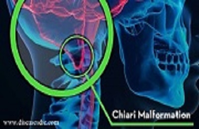
Chiari malformation is a congenital defective condition of the skull’s structure, in which cerebellum and the brain stem have penetrated into the foramen magnum, an opening at the base of the skull. Chiari is a term named after a pathologist Hans Chiari, who first described the Chiari malformation. The other terms are also used to refer Chiari malformation such as Arnold-Chiari malformation (this name usually refers specifically to Type II Chiari malformation) and hindbrain hernia.
Pathophysiology
Generally, the parts of the brain stem and cerebellum are located in the gap between the foramen magnum. In some cases, this gap in between the foramen magnum is abnormal in its size, thus the brain stem and cerebellum protruded or extended towards the top of the spinal canal through this space. This abnormal condition creates a pressure at the base of the brain and blocks the flow of cerebrospinal fluid (CSF) between the brain and spinal cord. Such problems affect the functions done by those parts of the brain.
Types of Chiari malformation
There are about four types of Chiari malformation as follows:
Type 1: Normally the spinal cord only passes into the foramen magnum, but in type 1 the lower part of the cerebellum (cerebellar tonsils) is extended till the foramen magnum. Type 1 is the most common form of Chiari malformation and doesn’t show any symptoms. It is usually noticed eventually when examining any other health issues during adulthood.
Type 2: It is also called as “classic” Chiari malformation or Arnold-Chiari malformation. Type 2 has severe symptoms more than type 1 and appears only during childhood period. In this type cerebellum and tissues of the brain stem protruded into the foramen magnum and also the nerve tissue between the two halves of the cerebellum is partially formed or absent. This type also associated with other conditions such as spina bifida and hydrocephalus.
Type 3: Type 3 is a very rare and serious form of Chiari malformation in which the cerebellum and the brain stem protrude through an abnormal opening at the back of the skull. Along with these protruded parts, the covering membranes of the brain and spinal cord also stick out. Symptoms usually appear during infancy and cause life-threatening effects in their futures. Infants with type 3 shows may show other neurological defects such as seizures, delay in physical and mental actions.
Type 4: Type 3 is different from other types because here the defect is with the development of the cerebellum. An underdeveloped or incompletely formed cerebellum (cerebellar hypoplasia) is located in its native place and some portions of the skull and spinal cord are exposed. This is a very rare form of Chiari malformation.
Causes and risk factors for Chiari malformation
The exact cause of CM is not known, but it tends to be developed from the birth. Some of the predicted causes are as follows.
- Chiari malformations are generally a reason for structural defects in the skull parts and spinal canal.
- These defects are developed when the genetic mutations occur during fetal development in its mother’s womb.
- Malnourished maternal diet lacks nutrient supplements to the fetus, thus causing congenital malformations during the fetal development.
- Chiari malformation also exists when the huge amount of cerebrospinal fluid is drained out as a result of injury, infection and toxic environmental exposure.
- CM also developed due to other conditions such as a tethered spinal cord, hydrocephalus, and in rare cases due to the tumor in the brain.
- Sometimes CM also due to the inherited mutated gene from their family members. However, the risk of passing altered genes from the family is very rare.
- Consuming illegal drugs and alcohol during pregnancy can also pose a reason for CM.
Incidence
Chiari malformations have no population studies on incidence and prevalence. From the clinical wise prevalence was estimated between 0.1 and 0.5%. But 0.77% as higher results of CM has resulted from the widespread use of MRI at a tertiary care center over a period of 3.5 years. From this 14 % were shown asymptomatic CM. The age distribution is majorly diagnosed in the late childhood period.
Symptoms
Many people do not have any symptoms. One of the common symptoms is a headache, especially during coughing, sneezing or straining. Other symptoms include:
- Difficulty while swallowing or speaking
- Scoliosis
- Balance or hearing problems
- Vomiting
- Insomnia
- Dizziness
- Numbness and muscle weakness
- Neck pain
- Poor coordination of hands and fine motor skills
- Depression
- Tinnitus (ringing or buzzing in the ears)
For some people, symptoms may vary depending upon the compression of the tissue and nerves and the build-up of pressure by cerebral spinal fluid (CSF).
Children with Chiari malformation may have difficulty in swallowing, being annoyed while feeding, a weak cry, excessive drooling, arm weakness, stiff neck, delay in development, and inability to gain weight.
Complications
In some people Chiari malformation may become serious disorder and causes complication such as follows:
Hydrocephalus: The excess fluid is filled in the brain and cause heavy pressure to the brain. The shunt tube is placed to drain the fluid.
Syringomyelia: A cyst (Syrinx) or cavity is formed in the spinal column. This may lead to serious discomforts for the usual head movement activity.
Tethered cord syndrome: In this condition, your spinal cord attaches to your spine and causes your spinal cord to stretch. This can cause serious nerve and muscle damage in your lower body.
Spina bifida: It is the condition in which your spinal cord or its covering isn’t fully developed, may occur in Chiari malformation, a part of the spinal cord is exposed out which cause serious paralysis. People with Chiari malformation type II usually have a form of spina bifida called myelomeningocele.
Diagnosis and test
Some of the tests and diagnosis for Chiari malformation include:
Magnetic resonance imaging scan (MRI): MRI image will provide the accurate view of the brain, cerebellum and spinal cord, it is good at providing and defining the extent of malformations. It can also evaluate the extent of fluid blockage and neural movement at the foramen magnum using CSF flow studies. MRI provides more information than CT scan about the back of the brain and spinal cord. It is the usual test preferred mostly for brain damages.
Computed tomography scan (CT): CT scan can be carried out to find the blockage and to define the size of the cerebral ventricles. It is useful for evaluating bony abnormalities at the bottom of the skull and cervical canal.
Swallowing study: Internal swallowing process is determined by fine any abnormalities abnormality suggestive of lower brainstem dysfunction by fluoroscopy (X-Rays).
Brainstem auditory evoked potential (BAER): To examine the function of hearing apparatus and brainstorm connections, an electrical test is carried out. It can also use to determine whether the brainstorm is working properly or not.
Myelogram: An X-ray of the spinal canal following injection of a contrast material into the CSF space; can show pressure on the spinal cord or nerves due to malformations. This test is performed less frequently now.
Somatosensory evoked potentials (SSEP): Sensation nerves are tested electrically, which gives some information about peripheral nerve, brain function, and spinal cord.
Sleep study: During sleep time breathing, snoring, oxygenation, and seizure activity are monitored to determine the sleep apnea.
Treatment and medications
Treatment is depending on the severity of the condition. If you don’t have symptoms your doctor will recommend regular imaging examinations. If you have primary symptoms like a headache, then your doctor may recommend pain medications.
Surgery to reduce pressure:
- Mostly Chiari malformation is treated with surgery, the main goal of the treatment is to stabilize your symptoms and to stop the changes in the anatomy of your brain
- This surgery can reduce the cerebellum pressure on your spinal cord and It restores the normal flow of spinal fluid
- Posterior fossa decompression is the most common surgery for Chiari malformation. A small portion of bone is removed in the back of your skull and relieving pressure by giving space to your brain
Posterior fossa decompression surgery
- In most of the cases, the brain covering called the dura mater is opened and a patch sewn is placed to enlarge the covering and to provide space to your brain.
- Sometime your doctor will remove a small portion of the spinal column to relieve pressure on your spinal cord and allow more space for the spinal cord
- The technique of the surgery may vary based on the fluid-filled cavity (Syrinx) present or fluid in your brain (hydrocephalus). If you have fluid in brain, the shunt tube is used to drain the extra fluids and relieves pressure in the brain
Prevention of chiari malformation
Chiari malformation is a congenital anomaly, and no method of prevention is known.

