Introduction
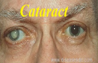
Cataract is a very common eye condition. As you get older the lens inside your eye gradually changes and becomes less transparent (clear). A lens that has turned misty, or cloudy, is said to have a cataract. Over time a cataract can get worse, gradually making your vision mistier. A cataract can occur in either or both eyes. It cannot spread from one eye to the other.
An eye with Cataract
Structure of Lens
Structure of an eye lens
The lens is a clear part of the eye that helps to focus light, or an image, on the retina. The retina is the light-sensitive tissue at the back of the eye. In a normal eye, light passes through the transparent lens to the retina. Once it reaches the retina, light is changed into nerve signals that are sent to the brain. The lens must be clear for the retina to receive a sharp image. If the lens is cloudy from a cataract, the image you see will be blurred.
Types of Cataracts
- Cataracts affecting the center of the lens (nuclear cataracts). A nuclear cataract may at first cause more nearsightedness or even a temporary improvement in your reading vision. But with time, the lens gradually turns more densely yellow and further clouds your vision.
- Cataracts that affect the edges of the lens (cortical cataracts). A cortical cataract begins as whitish, wedge-shaped opacities or streaks on the outer edge of the lens cortex. As it slowly progresses, the streaks extend to the center and interfere with light passing through the center of the lens.
- Cataracts that affect the back of the lens (posterior subcapsular cataracts). A posterior subcapsular cataract starts as a small, opaque area that usually forms near the back of the lens, right in the path of light. A posterior subcapsular cataract often interferes with your reading vision, reduces your vision in bright light, and causes glare or halos around lights at night.
- Cataracts you’re born with (congenital cataracts). Some people are born with cataracts or develop them during childhood. These cataracts may be genetic, or associated with an intrauterine infection or trauma.
History behind cataract
- Cataracts have been known to mankind for centuries. The word cataract comes from the Latin word “cataracta” meaning waterfall, with the condition possibly therefore named after the white appearance of rapidly running water.
- Couching is the earliest surgical procedure to be used in the treatment of cataracts. The procedure was first described by Maharshi Sushruta, an ancient Indian surgeon, in his treatise called the “Sushruta Samhita, Uttar Tantra” dating back to 800 B.C.E. The couching procedure, in which a needle is used to force the lens to towards the back of the eye. The eye would then be soaked with clarified butter and bandaged.
- Couching was eventually replaced by cataract extraction surgery, which used a suction device to remove the lens. Jacques Daviel (1696–1762), a French ophthalmologist, was one of the first European physicians to successfully extract cataracts from the eye, with his first operation dated to April 8, 1747.
- The pioneer of artificial intraocular lens transplant surgery was the English ophthalmologist Sir Nicholas Harold Lloyd Ridley who performed the first procedure in 1949.
- Phacoemulsification was introduced by ophthalmologist Charles D. Kelman in 1967.
Prevalence in world wide
According to the latest assessment, cataract is responsible for 51% of world blindness, which represents about 20 million people (2010). Although cataracts can be surgically removed, in many countries barriers exist that prevent patients to access surgery. Cataract remains the leading cause of blindness. As people in the world live longer, the number of people with cataract is anticipated to grow. Cataract is also an important cause of low vision in both developed and developing countries.
Causes behind the development of cataract
Cataracts can be caused by a number of things, but by far the most common reason is growing older. Most people over the age of 65 have some changes in their lens and most of us will develop a cataract in time. Apart from getting older, the other common causes of cataract include:
- Diabetes
- Trauma
- Medications, such as steroids
- Eye surgery for other eye conditions
- Frequent X-Ray or radiation transmission into head
In general, the reason why you have developed a cataract will not affect the way it is removed. Most cataracts are caused by natural changes in your lens, which happen as you get older. However, the following factors may be involved in cataract development:
- Tobacco smoking
- Lifelong exposure to sunlight
- Having a poor diet lacking antioxidant vitamins
Risk factors
Besides aging, other cataract risk factors include:
- Having parents, brothers, sisters, or other family members who have cataracts
- Having certain medical problems, such as diabetes
- Having had an eye injury, eye surgery, or radiation treatments on your upper body
- Having spent a lot of time in the sun, especially without sunglasses that protect your eyes from damaging ultraviolet (UV) rays
- People who have had the vitreous gel removed from their eye (vitrectomy) have an increased risk of cataracts.
Signs and symptoms
Signs and symptoms of cataracts include:
- Clouded, blurred or dim vision
- Increasing difficulty with vision at night
- Sensitivity to light and glare
- Need for brighter light for reading and other activities
- Seeing “halos” around lights
- Frequent changes in eyeglass or contact lens prescription
- Fading or yellowing of colors
- Double vision in a single eye
At first, the cloudiness in your vision caused by a cataract may affect only a small part of the eye’s lens and you may be unaware of any vision loss. As the cataract grows larger, it clouds more of your lens and distorts the light passing through the lens. This may lead to more noticeable symptoms.
Diagnosis and testing
Slit-lamp exam
Your ophthalmologist will examine your cornea, iris, lens and the other areas at the front of the eye. The special slit-lamp microscope makes it easier to spot abnormalities. A slit lamp allows your eye doctor to see the structures at the front of your eye under magnification.
The microscope is called a slit lamp because it uses an intense line of light, a slit, to illuminate your cornea, iris, lens, and the space between your iris and cornea. The slit allows your doctor to view these structures in small sections, which makes it easier to detect any tiny abnormalities.
Retinal exam
To prepare for a retinal exam, your eye doctor puts drops in your eyes to open your pupils wide (dilate). When your eye is dilated, the pupils are wide open so the doctor can more clearly see the back of the eye. Using the slit lamp, an ophthalmoscope or both, the doctor looks for signs of cataract. Your ophthalmologist will also look for glaucoma, and examine the retina and optic nerve.
Refraction and visual acuity test
This test assesses the sharpness and clarity of your vision. Each eye is tested individually for the ability to see letters of varying sizes. Using a chart or a viewing device with progressively smaller letters, your eye doctor determines if you have 20/20 vision or if your vision shows signs of impairment.
Treatment and Medications
Surgery
- Surgery to remove a cataract is the only way to get rid of a cataract. This surgery works well and helps people see better
- Cataract surgery involves removing the clouded lens and replacing it with a clear artificial lens. The artificial lens, called an intraocular lens (IOL), is positioned in the same place as your natural lens. It remains a permanent part of your eye.
- Most IOLs are made of silicone or acrylic. They are also coated with a special material to help protect your eyes from the sun’s harmful ultraviolet (UV) rays.
Days or weeks after surgery:
- You will have to use eye drops after surgery. Be sure to follow your doctor’s directions for using these drops.
- Avoid getting soap or water directly in the eye.
- Do not rub or press on your eye. Your ophthalmologist may ask you to wear eyeglasses or a shield to protect your eye.
- You will need to wear a protective eye shield when you sleep.
- Your ophthalmologist will talk with you about how active you can be soon after surgery. He or she will tell you when you can safely exercise, drive or do other activities again.
Medications for cataract
- Researchers have discovered that an organic compound called lanosterol can improve vision by dissolving the clumped proteins that form cataracts, said study lead author Dr. Kang Zhang, chief of ophthalmic genetics with the Shiley Eye Institute at the University of California, San Diego.
- Lanosterol eye drops could provide a cheaper and easier alternative for cataract treatment in many people, and perhaps prevent cataracts in someone at risk for developing them.
- Nepafenac Ophthalmic is a non-steroidal anti-inflammatory drug (NSAID), prescribed for eye pain, redness, and swelling in patients who are recovering from cataract surgery.
Complications after cataract surgery
Like any surgery, cataract surgery carries risks of problems or complications. Here are some of those risks:
- Eye infection.
- Bleeding in the eye.
- Ongoing swelling of the front of the eye or inside of the eye.
- Swelling of the retina (the nerve layer at the back of your eye).
- Detached retina (when the retina lifts up from the back of the eye).
- Damage to other parts of your eye.
- Pain that does not get better with over-the-counter medicine.
- Vision loss.
- The IOL implant may become dislocated, moving out of position.
Prevention of eye from cataract
There is no proven way to prevent cataracts. But certain lifestyle habits may help slow cataract development. These include:
- Not smoking.
- Wearing a hat or sunglasses when you are in the sun.
- Avoiding sunlamps and tanning booths.
- Eating healthy foods.
- Avoiding the use of steroid medicines when possible (some people need them).
- Keeping diabetes under control.

